Scedosporium apiospermum Sacc. ex Castell. & Chalm., Manual of tropical medicine (London): 1122 (1919)
MycoBank number: MB 432048;Index Fungorum number: IF 432048; Facesoffungi number: FoF 11704 Fig. x
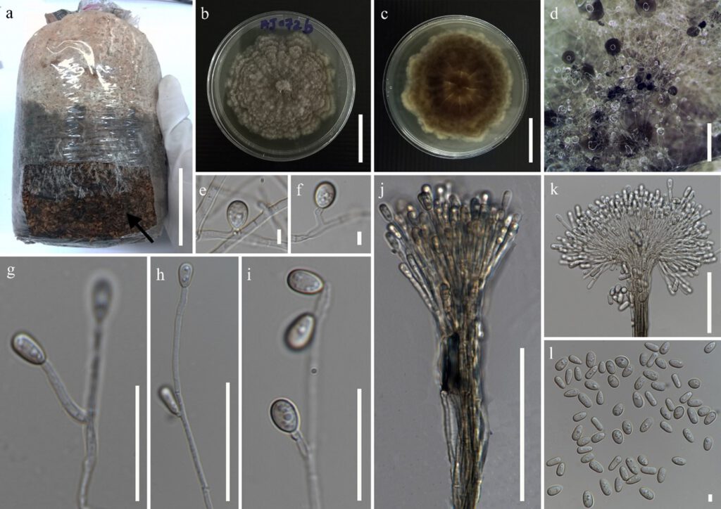
Growing on Oyster mushroom grow substrate. Sexual morph: Not observed. Asexual morph: on host, Mycelium black powdery mass. On PDA, hyphomycetous, Hyphae 1.5–3.5 μm (x̅ = 2.2 μm) wide, branched, septate, hyaline. Conidiophores solitary or synnematous, solitary conidiophores 75–128 × 1.8–3 μm (x̅ = 102.5 × 2.6 μm, n = 10) hyaline, branched, forms 1–3 conidiogenous cells at the end. synnematous conidiophores 132–352 × 8.2–17.5 μm (x̅ = 288.4 × 12.7 μm, n = 15) cylindrical stipe, erect, forms conidia at the end. Conidiogenous cells 4.8–25 × 1.2–2.5 μm (x̅ = 15.2 × 1.3 μm, n = 10) lateral or terminal formation on solitary conidiophores, hyaline, cylindrical. Conidia hyaline, guttulate, conidia arising from conidiogenous cells 6.2–8.2 × 2–4.5 μm (x̅ = 7.4 × 3.3 μm, n = 30) obvoid to ellipsoidal, conidia arising from synnematous conidiophores 6.4–9.2 × 1.7–3.5 μm (x̅ = 7.5 × 2.8 μm, n = 35) cylindrical or claviform with a truncate base, conidia from undifferentiated hyphae 6–7.5 × 5.2–6.6 μm (x̅ = 6.4 × 5.2 μm, n = 20) sessile, globose to subglobose.
Culture characteristics: colonies on PDA attaining 65–75 mm diameter after 14 days at 25 °C, cottony, dense, lanose, light gray to dark grey, margins fimbriate and irregular; reverse dark grey to brown, grey to white margins.
Material examined: Thailand, Phayao province, growing on the oyster mushroom growing substrate, 06 June 2020, AJ. Gajanayake, AJ 072 (inactive dry culture, MFLU 22-xxxx, new host record), living culture, MFLUCC 22-xxxx.
GenBank numbers: ITS = xxxxx, tub2 = xxxxx.
Notes: Our isolate MFLUCC 22-xxxx, clusters within Scedosporium apiospermum with a boostrap support of 90% ML, 0.97 BYPP. Furthermore, the morphology of our isolate MFLUCC 22-xxxx resembles the original description and illustrations for Scedosporium apiospermum by Saccarado in Castellani and Chalmers (1919) and the description and morphological illustrations by Gilgado et al. (2010). However, there are size differences of conidiophores, conidiogenous cells and conidia, when we compare our strain with the strains described in Castellani and Chalmers (1919) and Gilgado et al. (2010). The reason for this may be the differences in the media in which the colonies were grown.
Scedosporium apiospermum has been mainly identified as an opportunistic clinical pathogen relevant to many infections which commonly occur in the bones, central nervous system, lungs, paranasal sinuses, skin and soft tissues (Shinohara et al. 2009; Goldman et al. 2016). Seaphueek et al. (2017) isolated 21 fungal species from spent mushroom substrate of Pleurotus sp. collected from mushroom farms in southern Thailand. Among those 21 fungal species there were, Alternaria spp., Aspergillus spp., Chaetomium sp., Cunninghamella spp., Fusarium spp., Lasiodiplodia sp., Neurospora sp., Penicillium spp., Rhizoctonia sp. and Trichoderma spp. (Seaphueek et al. 2017). Suada et al. (2015) have reported Aspergillus spp., Fusarium spp., Gliocladium sp., Mucor spp., Neurospora spp., Paecilomyces sp., Penicillium spp., Pythium sp., Stachybotrys spp. and Trichoderma spp. as contaminants from oyster mushroom growing substrate. According to best our knowledge this is the first report of Scedosporium apiospermum as a contaminant of oyster mushroom growing substrate.

Fig. x Scedosporium apiospermum (MFLU 22-xxx) a. Contaminated oyster mushroom grow substrate. b,c. Colony on PDA. d. Sporulation of the colony on PDA. e-i. Conidial attachments and conidiogenous cells. j,k. Synnema with conidia. l. Conidia. Scale bars: a = 5 cm, b,c = 30 mm, d = 200 μm, e,f,l = 4 μm, g = 20 μm, h = 35 μm, i = 25 μm, j = 65 μm, k = 40 μm.
