Sajamaea mycophila Flakus, Piątek & Rodr. Flakus, sp. nov. (Fig. 4)
MycoBank number: MB 832197; Index Fungorum number: IF 832197; Facesoffungi number: FoF;
Etymology: The epithet name mycophila refers to the occurrence of this fungus on other fungus.
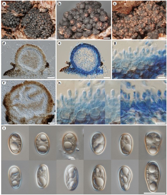
Description: Mycoparasitic on Paraleptosphaeria polylepidis, causing moderate damages of host ascomata. Conidiomata pycnidial, as pale brown galls on host surface, 150–300 μm wide, 120–300 μm high, first erumpent through the outermost layer of host peridium, later almost sessile on pseudothecial clusters of host, uniloculate to multi-loculate, solitary to aggregated, subglobose to irregular in shape, pale brown; better seen when wet. Peridium 20–30 μm wide, pale to dark brown, composed of about 3–8 layers of cells with walls up to 2 μm wide, cells isodiametric to slightly elongate, textura angularis. Conidiophores reduced to conidiogenous cells. Conidiogenous cells 5–11 μm high, 4–8 μm wide, enteroblastic, phialidic, smooth-walled, hyaline, with a small collar, densely outlining inner surfaces of pycnidia. Conidia 9–13 × 5.5–7.5 μm, pale brown, broadly ellipsoidal, sometimes slightly narrower at one point, with rounded ends, aseptate, smooth, thin-walled, guttulate. Sexual state unknown.
Specimen examined: Bolivia, Oruro, Sajama National Park (=Parque Nacional Sajama), W slope of Nevado Sajama, closely located to the permanent monitoring plot “Cohiri,” 19K 512102, 8002927, elev. ca. 4425 m, parasitic on Paraleptosphaeria polylepidis growing on Polylepis tarapacana, 15 Oct. 2016, A.N. Palabral-Aguilera (APA- 2999), C. López & M.I. Gómez (LPB 0003512 – holotype, KRAM F-59659 – isotype).

Fig. 4 Sajamaea mycophila (KRAM F-59659). a–c Habit of pale brown conidiomata growing on ascomata of Paraleptosphaeria polylepidis. d–e Longitudinal section of uniloculate conidioma erumpent through the outermost layer of host peridium (e mounted in LPCB). f Longitudinal section of multi-loculate conidioma showing layers of conidiogenous cells (hyaline areas) and conidial masses (brown areas). g Section through paraplectenchymatous peridium showing conidiogenous cells mounted in LPCB. h Conidiogenous cells and young conidia mounted in LPCB. i Conidia. Scale bars: a 1000 μm, b–c 500 μm, d–f 50 μm, g–i 10 μm
