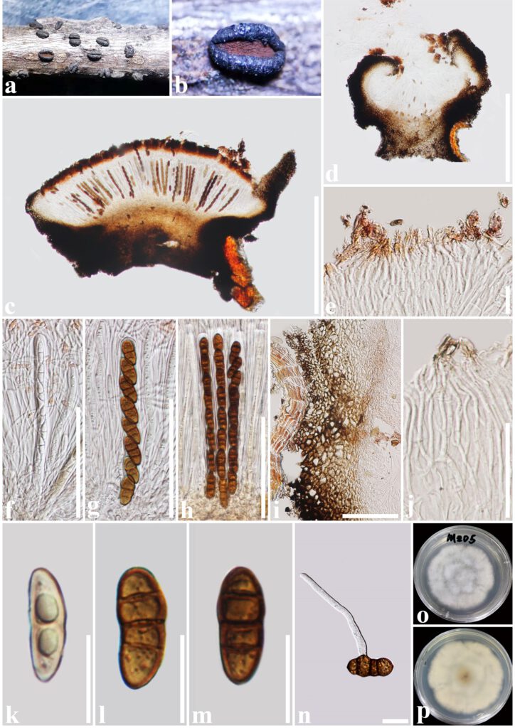
Rhytidhysteron mengziense T.Y. Du and Tibpromma sp. nov. (Figure 3)
MycoBank number: MB; Index Fungorum number: IF; Facesoffungi number: FoF 12959;
Etymology: Named after the region, where this sample was collected, Mengzi.
Holotype: HKAS 122699
Saprobic on decaying twigs of Crataegus scabrifolia (Rosaceae). Sexual morph: Ascomata 1000–1600 μm long × 800–1000 μm wide × 400–700 μm high (x̅ = 1400 × 910 × 640 μm, n = 5), hysterothecial, solitary to aggregated, mostly solitary, semi-immersed to superficial, navicular, black, apothecioid, smooth, perpendicularly striate, elongate and depressed, compressed at apex, opening by a longitudinal slit, reddish-brown at the center. Exciple 60–135 μm wide, composed of outer layer brown to black, thick-walled cells of textura angularis, and inner layer light brown, thin-walled cells of textura prismatica. Hamathecium comprising 1–2.5 μm wide, dense, hyaline, septate, branched, cellular pseudoparaphyses, forming a reddish-brown to brown epithecium above asci when mounted in water, becoming purple epithecium above the asci when mounted in 10% KOH and turns hyaline after 30 seconds, while appendages turn dark brown. Asci 150–176(–182) μm × 10–14(–16.3) μm (x̅= 164.5 × 13 μm, n = 20), 8-spored, bitunicate, cylindrical, with short pedicel, rounded at the apex, with an ocular chamber, J–, always fused with hamathecium. Ascospores (22.5–)24.5–27.5(–29) μm × 10.5–12.5 μm (x̅= 27 × 12 μm, n = 30), slightly overlapping, uni-seriate, slightly overlapping, hyaline when immature, becoming reddish-brown to brown when mature, ellipsoidal to fusoid, straight or curved, rounded to slightly pointed at both ends, 3-septate, guttulate, rough-walled, without the mucilaginous sheath. Asexual morph: Undetermined.
Culture characteristics: Ascospores germinating on PDA within 24 h and germ tubes produced from one or both ends. Colonies on PDA reached a 6 cm diameter after one week at 28°C. The colony flossy, velvety, circular, slightly raised, with an undulated edge, white aerial hyphae on the forward and cream white in reverse.
Material examined: China, Yunnan Province, Honghe Prefecture, Mengzi City, on decaying wood of Crataegus scabrifolia (Rosaceae), 21 May 2020, S. Tibpromma, MZD5 (holotype, HKAS 122699, ex-type living culture, MFLUCC 21-0490, living culture, MFLUCC 21-0491).
Notes: Rhytidhysteron mengziense was closely related to Rhytidhysteron camporesii Ekanayaka & K.D. Hyde, formed a well separate branch, but with low bootstrap support. However, R. mengziense is distinct from R. camporesii in having exciple cells of textura angularis to prismatica, and rough-walled of ascospores, while R. camporesii has exciple cells of textura globulosa to angularis, and smooth-walled of ascospores (Hyde et al. 2020b). In addition, the ascomata and ascospores size of R. mengziense are larger than those of R. camporesii (ascomata: 1400 × 640 μm vs 1002.4 × 570.1 µm, ascospores: 27 × 12 μm vs 26.1 × 10.4 µm) (Hyde et al. 2020b). Moreover, according to the comparison results of different gene fragments, R. mengziense is different from R. camporesii in ITS gene (10/651 bp, 1.54%). Therefore, in this study, R. mengziense is introduced as a new species.

Figure 3. Rhytidhysteron mengziense (HKAS 122699, holotype). a, b Appearance of hysterothecia on the host. c, d Vertical section through hysterothecium. e Epithecium mounted in water. f-h Asci. i Exciple. j Pseudoparaphyses. k-m Ascospores. n. A germinating ascospore. o, p. Colony on PDA medium (after one week). Scale bars: c, d = 500 μm, e, k-n = 20 μm, f-i = 100 μm, j = 50 μm.
