Rhodofomitopsis pseudofeei B.K. Cui & Shun Liu, sp. nov.
Index Fungorum number: IF 829661; MycoBank number: MB 829661; Facesoffungi number: FoF 06041; Figs. 102, 103
Etymology: Referring to the species being similar to Rhodofomitopsis feei (Fr.) B.K. Cui, M.L. Han & Y.C. Dai in morphology.
Holotype: Cui 16794 (BJFC).
Basidiocarps annual, pileate, sessile, solitary or imbricate, easily separable from the substrate, without odor or taste, corky when fresh, becoming hard corky and light in weight when dry. Pilei applanate, semicircular to flabelliform, projecting up to 4.2 cm, 8.7 cm wide and 2.5 cm thick at base. Pileal surface greyish brown to fuscous at base and cream to honey yellow toward the margin, glabrous to slightly velutinate, azonate, slightly sulcate or not; margin obtuse to acute. Pore surface clay buff to clay pink when dry; sterile margin distinct, pale mouse grey to greyish brown, up to 4 mm wide; pores round and small, but somewhat variable, mostly 5–7 per mm and almost invisible to the naked eye; dissepiments thick, entire. Context buff to reddish brown or cinnamon brown, corky, up to 15 mm thick. Tubes concolorous with pore surface, hard corky, up to 2 mm long. Hyphal system dimitic; generative hyphae bearing clamp connections; skeletal hyphae IKI–, CB–; tissues becoming blackish brown in KOH. Context generative hyphae hyaline, thin-walled, occasionally branched, 1.7–3.8 µm diam; skeletal hyphae dominant, hyaline to pale yellowish, thick-walled with a wide to narrow lumen, frequently simple-septate, occasionally branched, interwoven, 2–5.5 µm diam. Trama generative hyphae hyaline, thin-walled, occasionally branched, 1.8–2.6 µm diam; skeletal hyphae dominant, hyaline to pale yellowish, thick-walled with a wide to narrow lumen, frequently simple-septate, occasionally branched, interwoven, 1.5–4 µm diam. Cystidia absent, but fusoid cystidioles occasionally present, hyaline, thin-walled, 12–23×3–5 µm. Basidia clavate, bearing four sterigmata and a basal clamp connection, 12–23×4–6.5 µm; basidioles dominant, in shape similar to basidia, but smaller. Basidiospores cylindrical, sometimes curved, hyaline, thin-walled, smooth, IKI–, CB–, (5.2–)5.9–7(–8.4) ×(2–)2.5– 3.2(–3.3) µm, L=6.57 µm, W =2.87 µm, Q =2. 19-2.32 (n=90/3).
Material examined: AUSTRALIA, Queensland, Cairns, nearby Mount Whitfield Conservation Park, on fallen angiosperm trunk, 7 May 2018, Cui 16794 (BJFC, holotype); on fallen angiosperm trunk, 7 May 2018, Cui 16762 (BJFC); on fallen angiosperm trunk, 7 May 2018, Cui 16803 (BJFC); on fallen angiosperm trunk, 7 May 2018, Cui 16807 (BJFC).
GenBank numbers: ITS: MK461952, MK461951, MK461953, MK461954; LSU: MK461956, MK461955, MK461957, MK461958; mtSSU: MK461960, MK461959, MK461961, MK461962; nSSU: MK461964, MK461963, MK461965, MK461966; RPB2: MK463984, MK463983, TEF1: MK463986, MK463985, MK463987.
Notes: Rhodofomitopsis pseudofeei was found from tropical areas of Australia. Morphologically, it is similar to R. feei by the pileate and pinkish basidiocarps (Gilbertson and Ryvarden 1986). However, the latter species has smaller basidiospores (5–6.5×2–3 µm), and lacks cystidioles (Han and Cui 2015). Phylogenetically, R. pseudofeei is distinct from R. feei (Fig. 101). Fomitopsis subfeei B.K. Cui & M.L. Han was recently described from China (Han and Cui 2015) and transferred to Rhodofomes Kotl. & Pouzar as a new combination: Rhodofomes subfeei (B.K. Cui & M.L. Han) B.K. Cui, M.L. Han & Y.C. Dai (Han et al. 2016), which also has pileate and pinkish basidiocarps and may be confused with Rhodofomitopsis pseudofeei, but it is distinguished by its bigger pores (4–6 per mm) and smaller basidiospores (4–5×1.9–2.4 mm, Han and Cui 2015).
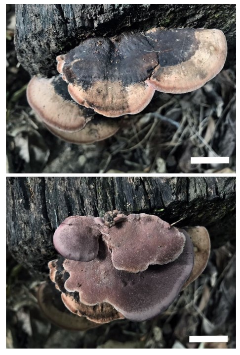
Fig. 102 Basidiocarps of Rhdofomitopsis pseudofeei (Cui 16794, holotype). Scale bar:=2 cm
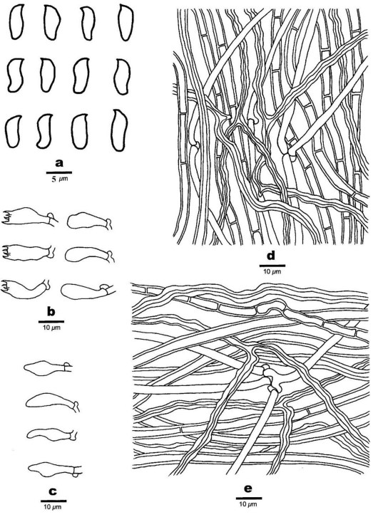
Fig. 103 Microscopic structures of Rhdofomitopsis pseudofeei (Cui 16794, holotype). a Basidiospores. b Basidia and basidioles. c Basidioles. d Hyphae form trama. e Hyphae from context