Rhizophydites matryoshkae M. Krings et C.J. Harper, sp. nov.
MycoBank number: MB 834568; Index Fungorum number: IF 834568; Facesoffungi number: FoF;
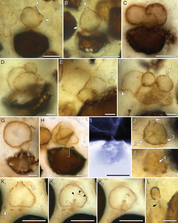
Diagnosis. Zoosporangia smooth walled, sessile, usually spheroidal, less than 35 mm in diameter but may also be drop shaped, rhomboidal, obpyriform, or somewhat elongate to spindle shaped; one to several generations of zoosporangia of decreasing size may occur one inside another or with large numbers of spheroidal or pyriform miniature zoosporangia in the old zoosporangium; one to four discharge openings, prominent, papilla-like or short tube–like, up to 5 mm high; miniature zoosporangia usually spheroidal, less than 10 mm in diameter, with a single distal discharge papilla or short tube; encysted zoospores spheroidal to ovoid, 2.5–4.5 (–5.5) mm in diameter, sessile or pedicellate, often present on outer surfaces of zoosporangia; rhizoidal system originating from base of zoosporangium or, if present, subsporangial swelling, extending into host lumen; hosts are spores of early land plants Horneophyton lignieri and Aglaophyton majus.
Holotype. Specimen in figure 2B from slide SNSB-BSPG 1964 XX 99, SNSB-BSPG, Munich, Germany.
Collection locality. Rhynie, Aberdeenshire, Scotland, National Grid Reference NJ 494276 (lat. 57720009.9700N, long. 0027 50031.8300W).
Stratigraphic position. Dryden Flags Formation.
Age. Early Devonian; Pragianearliest Emsian (see Wellman 2006, 2017; Wellman et al. 2006), 411:5 5 1:3 Ma (Parry et al. 2011), 407:1 5 2:2 Ma (Mark et al. 2011).
Remarks. Rhizophydites matryoshkae is based on considerable information on the morphology, together with specific developmental details, that renders the form distinct, readily recognizable, and distinguishable from other Rhynie chert fungi; makes the formulation of a diagnosis containing a sufficiently large set of diagnostic characters possible; and enables taxonomic assignment (see “Discussion” below). However, because of the geologic age and given the need for molecular data in fungal taxonomy today, we refrain from assigning the fossil to any one of the present-day chytrid genera and species but rather propose a new fossil taxon for the form. We also include in R. matryoshkae several unnamed specimens of A. majus figured by Taylor et al. (1992b: figs. 19, 23, 24) because they are not distinguishable morphologically from the fossils described in this study. All specimens are interpreted as belonging to a single fossil species, albeit with the caveat that they might represent several biological species that are impossible to distinguish on the basis of the material at hand.
Etymology. The genus name acknowledges the morphological congruence that exists between the fossil and the present-day genus Rhizophydium (Rhizophydiales; Chytridiomycota), in particular Rhizophydium proliferum J.S. Knox et R.A. Paterson; the endingites is used to designate a fossil taxon, as recommended by Pirozynski and Weresub (1979). The epithet is proposed because some of the specimens are reminiscent of Russian matryoshka dolls, which are the sets of wooden dolls of decreasing size placed one inside another.

Fig. 2 Morphology of Rhizophydites matryoshkae. All specimens are from slide SNSB-BSPG 1964 XX 99 unless indicated otherwise. A, B, Mature thalli with short discharge tubes (arrows). C, Spheroidal zoosporangia arising from a common site in the host wall. Slide SNSB-BSPG 2013 V 27. D–F, Zoosporangia in different phases of development, some arising from a host spore, others from the surfaces of adjacent zoosporangia; note the miniature zoosporangium attached via a rhizoidal axis in E (arrow) and the discharge papillae or short tubes (arrows) in F. G, Spheroidal zoo- sporangium with subsporangial swelling. Slide SNSB-BSPG 2013 V 27. H, Bundle of zoosporangia emerging from the host spore. Slide SNSB-BSPG 2013 V 27. I, Detail of H (bracketed area) in inverted light, focusing on the rhizoidal system. J1, J2, Zoosporangium with four discharge papillae (denoted as 1–4); note the several encysted zoospores on the outer zoosporangium surface in J2. K1–K3, Different focal planes of a zoosporangium with one discharge tube. The arrows in K1 indicate encysted zoospores in the immediate vicinity; the arrow in K2 shows an encysted zoospore in the lumen, and the arrowheads point at a rhizoidal axis extending into the host lumen from an encysted zoospore attached to the outer surface. Note what appears to be a developing miniature zoosporangium inside the parent zoosporangium in K3. L, Encysted zoospore with a developing rhizoidal axis (arrow). Scale bars p 25 mm (A–C, K1–K3), 10 mm (D–J2), 5 mm (L).
