Rectifusarium ventricosum (Appel & Wollenw.) L. Lombard & Crous, in Lombard, van der Merwe, Groenewald & Crous, Stud. Mycol. 80: 229 (2015)
MycoBank number: MB 810253; Index Fungorum number: IF 810253; Facesoffungi number: FoF 10991; Fig. 79
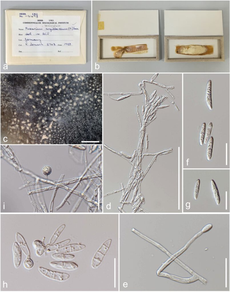
Isolated from soil. Asexual morph: Hyphomycetous. Colonies white, effuse, hairy or velvety. Mycelium superficial, comprising branched, septate, hyaline hyphae. Conidiophores 75–90 × 2–5 µm (x̅ = 81.1 × 3.5 µm, n = 5), mononematous, originating laterally from hyphae, generally unbranched, sometimes branched at the apex, septate, simple, straight to flexuous. Conidiogenous cells 35–70 × 2–4 µm (x̅ = 45.1 × 3.1 µm, n = 5), terminal, monophialidic, cylindrical, slightly tapering towards the apex, periclinal thickening visible, apex with minute collarettes, hyaline, smooth-walled. Macroconidia 19–30 × 4–6.5 µm (x̅ = 23.1 × 5.3 µm, n = 25), 1–3-septate, fusiform to ellispoidal, straight or slightly curved, rounded at each ends or basal end of cells slightly flattened, hyaline, smooth-walled. Chlamydospores 8–9.5 × 7.5–9 µm (x̅ = 8.7 × 8.5 µm, n = 5), arising terminally or laterally, hyaline to subhyaline, globose, thick-walled, verruculose, solitary or forming chains or developing directly from macroconidia. Microconidia not observed. Sexual morph: See Booth (1971).
Material examined: GERMANY, isolated from soil, 1958, K. Domsch, IMI 112498.

Fig. 79 Rectifusarium ventricosum (IMI 112498,) a Herbarium packet b Dried culture herbarium c Close-up of colony d, e Hyphae, conidiophores and conidiogenous cells f, g Macroconidia h Macroconidia and chlamydospores developing from macroconidia i Chlamydospore. Scale bars: c =2 mm, d = 100 µm, e–i = 20 µm.
