Pulmosphaeria archontophoenicis Joanne E. Taylor, K.D. Hyde & E.B.G. Jones, Sydowia 48(2): 256 (1996)
Index Fungorum number: IF 436688; MycoBank number: MB 436688; Facesoffungi number: FoF 01354
Saprobic on dead petioles of Archontophoenix alexandrae (F. Muell.) H. Wendl. & Drude. Sexual morph: Ascomata 570–740 × 110–200 µm (x̄ = 678 × 185 μm, n = 10), immersed in a poorly developed stroma, developing beneath the slightly raised areas comprising host cuticle and epidermal cell layers, occurring in pairs, with a central ostiole, cylindrical, with their long axis horizontal to the host surface, in cross-section 310–425 × 110–200 µm (x̄ = 376 × 187 μm, n = 10). Peridium composed of several cell layers, outwardly comprising light brown, angular cells, inwardly comprising hyaline, angular cells. Hamathecium comprising few, deliquescent paraphyses, less than 5 µm wide. Asci 210–330 × 23–36 µm (x̄ = 289 × 28 μm, n = 20), 8-spored, unitunicate, cylindrical, short pedicellate, apical apparatus not bluing in Melzer’s reagent, J-, indistinct, 5–9 × 5–6 µm (x̄ = 7.4 × 5.6 μm, n = 20). Ascospores 152–200 × 7.5–11 µm (x̄ = 187 × 9 μm, n = 20), crowded, unicellular, hyaline, filiform, apex obtuse, tapering to base, 1-septate, septum 45–62.5 µm (x̄ = 57 μm, n = 20) from the base, smooth-walled. Asexual morph: Undetermined.
Material examined – AUSTRALIA, Queensland, on dead petiole of Archontophoenix alexandrae (F. Muell.) H. Wendl. & Drude, 17 April 1995, J.E. Taylor and K.D. Hyde, BRIP 23234 holotype.
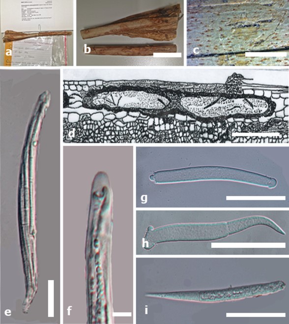
Fig. 1. Pulmosphaeria archontophoenicis (holotype) a Herbarium specimen details. b, c Stromata in wood. d Drawing of cross section of stromata showing ascomata encased in stromatal tissue. e Mature asci. f Asci with J- apical apparatus not bluing in Melzer’s reagent. g–i Ascospores. Scale bars: b = 5 mm, c = 1000 µm, d= 200 µm, e = 20 µm, f = 5 µm, g–i = 50 µm.