Pseudopithomyces maydicus (Sacc.) J.F. Li, Ariyawansa & K.D. Hyde.
Basionym – Clasterosporium maydicum Sacc., Nuovo G. bot. ital. 23(2): 213 (1916)
≡ Pithomyces maydicus (Sacc.) M.B. Ellis, Mycol. Pap. 76:15 (1960)
For other synonyms see Index Fungorum (2015)
Index Fungorum number: IF551395; Facesoffungi number: FoF00940; Fig. 2
Saprobic on leaves of Acoelorrhaphe wrightii. Sexual morph: Undetermined. Asexual morph: Colonies effuse, dark brown to black. Mycelium superficial or partly immersed in the substrate, composed of septate, branched, smooth, thinwalled, white hyphae. Conidiophores 4 – 6 × 2.5 – 3 μm (x̄ = 4.4 × 2.7 μm, n = 100), semi-micronematous, mononemous, closely packed, hyaline to subhyaline, thin-walled, aseptate, smooth, unbranched, flexuous. Conidiogenous cells 3.5 – 5 × 1.4 – 4 μm (x̄ = 3.6 × 2.2 μm, n = 100), monoblastic, integrated, terminal, determinate, hyaline, globose or subglobose. Conidia 13.5 – 24.5 × 5.5 – 8.5 μm (x̄ = 21.8 × 5.6 μm, n = 100), wide in the middle, acrogenous, muriform, 3 – 4 – septate, thin-walled, brown to dark brown, fusiform to pyriform, clavate.
Cultural characteristics – Conidia germinating on PDA within 14 h and germ tubes produced from apical cells. Colonies growing on PDA, hairy or cottony, white to dark grey, reaching 5 mm in 20 days at 25 °C, mycelium superficial, effuse, radially striate, with regular edge, hypha white to grey. Asexual spores and sexual spores not formed within 60 days.
Material examined – THAILAND, Chiang Rai Provice, Mae Fah Luang, on dead leaves of Acoelorrhaphe wrightii (Arecaceae), 26 Janurary 2014, J.F. Li, H-4e (MFLU 15- 1473, reference specimen designated here), living culture, MFLUCC 14-0391, KUM-15-0101.
Notes – Pseudopithomyces maydicus is characterised by cylindrical or slightly clavate, pale brown to dark brown, verrucose conidia with two to three transverse septa, rarely with one oblique or longitudinal septum and in its white to grayish yellow colonies on PDA. The characters of our collection are typical with that of Clasterosporium maydicum (Ellis 1960). Furthermore our phylogeny also concluded that the putative strains of Pse. maydicus (UTHSC 06 3954 and UTHSC 06 1549) clustered with our strain (Fig. 1).
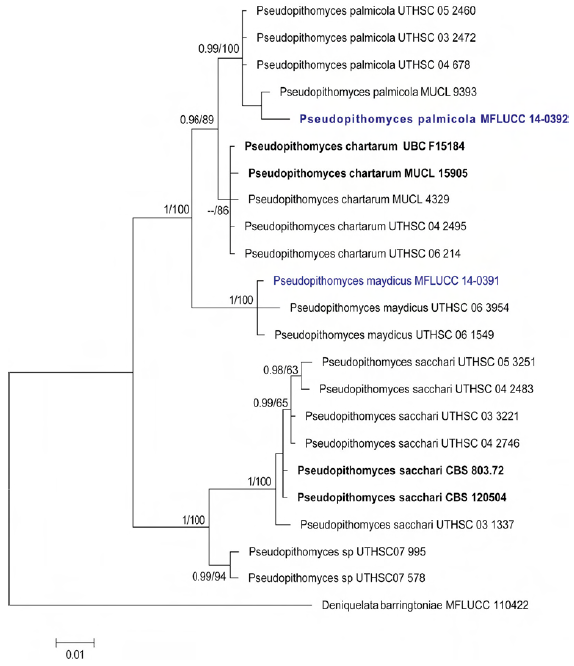
Fig. 1 Phylogram generated from Bayesian analysis based on ITS and LSU sequence data. Bayesian posterior probabilities (PP) greater than 0.90 are indicated above or below the nodes. The ex-type and reference strains are in bold, the new isolates are in blue. The tree is rooted with Deniquelata barringtoniae (MFLUCC 11-0422)
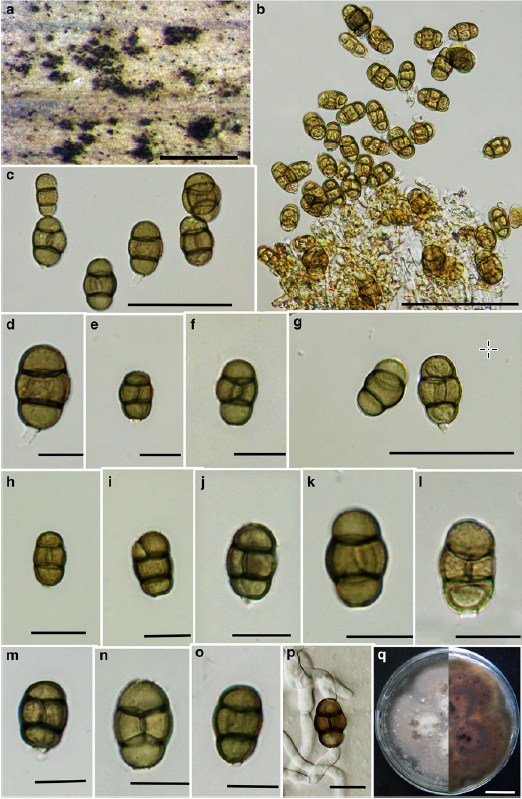
Fig. 2 Pseudopithomyces maydicus (reference specimen) a Colonies on palm stem b Conidiogenous cells and conidiophores bearing conidia c – o Conidia p Germinated conidium q Upper and lower view of colony. Scale bars: a = 500 μm, b = 100 μm, c, g = 50 μm, d – n = 20 μm, q = 3 cm.