Pseudoophiobolus urticicola Phookamsak, Wanas., Camporesi & K.D. Hyde, sp. nov. Index Fungorum number: IF553930
Etymology: The specifific epithet ‘‘urticicola’’ refers to the host Urtica
Holotype: MFLU 17-0933
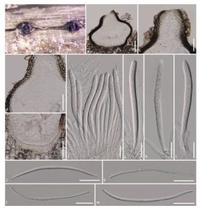
Saprobic on Urtica dioica L. Sexual morph: Ascomata 240–300 µm high (excluding papilla), 230–390 µm diam., dark brown to black, scattered, solitary to gregarious, semi immersed, subglobose to ampulliform, uni-loculate, glabrous, ostiolate, papillate. Papilla 95–170 µm high, 90– 185 µm diam., with rounded to truncate apex, composed of several layers of small, thickened, black, pseudoparenchymatous cells, arranged in a textura angularis, ostiole central, pore-like opening, with hyaline periphyses. Peridium 15–40 µm wide, slightly thicker at the base, thinner towards the apex, composed of several cell layers of flflattened, small, thick-walled, pseudoparenchymatous cells, arranged in textura angularis to textura prismatica, hyaline cells at the base, with dark brown to black cells at the sides towards the apex. Hamathecium composed of dense, 1.5–3 lm wide, filamentous, cellular pseudoparaphyses, distinctly septate, not constricted at the septa, anastomosing at the apex, embedded in a hyaline gelatinous matrix. Asci (107–)120–130(–138) x 8–10 µm (x = 124.4 x 9.1 µm, n = 30), 8-spored, bitunicate, fissitunicate, cylindrical to cylindric-clavate, short pedicellate, apically rounded, with well-developed ocular chamber. Ascospores (77–)80–100(–107) x 2–3 µm (x = 93.9 x 3.7 µm, n = 30), fasciculate, scolecosporous, subhyaline to pale yellowish at maturity, fifiliform, with rounded ends, tapered towards the end cells, swollen at the base of the 4th or 6th cell, 9–13-septate, not constricted at the septa, smooth-walled. Asexual morph: Undetermined.
Culture characteristics: Colonies on PDA reaching 28–30 mm diam. after 4 weeks at 25–30 C, medium dense, irregular, flattened, slightly raised at the middle, surface smooth, with pale pinkish turfs, and small, yellowish droplets, edge lobate, entire margin, velvety, colony from above, pale orangish at the magin, orangish brown to dark brown at the centre, radiating with paler concentric rings, colony from below, orangish-yellow at the margin, paler in the middle, orangish brown to dark brown at the centre, not producing pigmentation in agar.
Material examined: ITALY, Province of Arezzo [AR], near Croce di Pratomagno, on dead stem of Urtica dioica L. (Urticaceae), 29 July 2014, E. Camporesi, IT 2030 (MFLU 17-0933, holotype), ex-type living culture, KUMCC 17-0168.
Notes: Pseudoophiobolus urticicola is similar to Ophiobolus collapsus in having ascospores swollen at the 4th cell near the septum. However, the two species can be distinguished based on ascospore colour and septation. Pseudoophiobolus urticicola has subhyaline to pale yellowish ascospores, with 9–13 septa, whereas O. collapsus has yellowish ascospores with 7 septa (Shoemaker 1976). Multigene analyses showed that P. urticicola clusters with P. erythrosporus and also closely related to P. achilleae and P. rosae. The ITS pairwise comparison shows that P. urticicola differs from P. achilleae and P. rosae in eight and four base positions respectively. Pseudoophiobolus urticicola also differs from P. rosae and P. galii in seven bases and 19 base positions respectively, based on the TEF1-a gene pairwise comparisons.
Hosts: Urtica dioica L. (Urticaceae)
Distribution: Italy
FIG Pseudoophiobolus urticicola (MFLU 17-0933, holotype). a Appearance of ascomata on the host surface. b Section through ascoma. c Section through papillate. d, e Section through peridium. f Asci embedded in pseudoparaphyses. g–i Asci. j–m Ascospores. Scale bars: b = 100 µm, c–e = 50 µm, f–m = 20 µm

