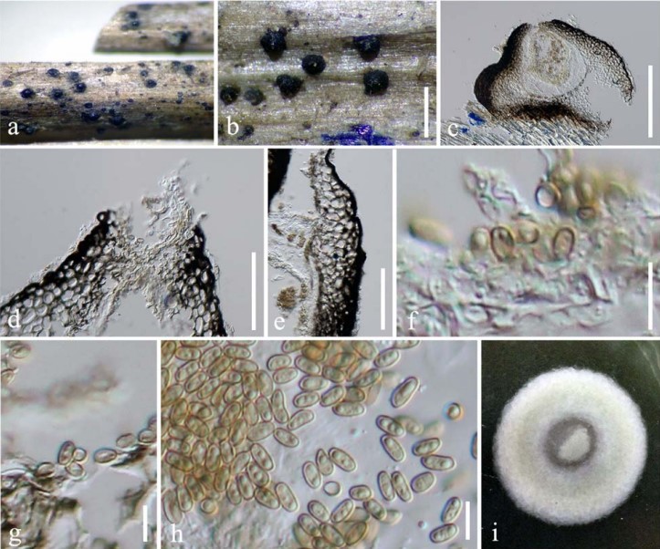
Pseudocyclothyriella clematidis (Phukhams. and K. D. Hyde) Phukhams. and Phookamsak, comb. nov., Figure 3
MycoBank number: MB 557442; Index Fungorum number: IF 557442; Facesoffungi number: FoF 09540;
Pseudocoleophoma clematidis Phukhams. and K. D. Hyde, in Phukhamsakda et al., Fungal Divers (102): 21 (2020).
Holotype Details: Italy, Arezzo Province, Badia Tega— Ortignano Raggiolo, on dead aerial branch of Clematis vitalba, March 9, 2013, E. Camporesi, IT 1110 (MFLU 16-0280), ex-type living culture, MFLUCC 17-2177.
Description and Illustration: See Phukhamsakda et al. (2020).
Ecology and Known Distribution: As a saprobe, Pseudocyclothyriella clematidis has contributions in the cycle of the material. To date, the species is just reported in Italy (Phukhamsakda et al., 2020).
Notes: Pseudocyclothyriella clematidis was previously treated in Pseudocoleophoma based on phylogenetic evidence (Phukhamsakda et al., 2020). Pseudocyclothyriella clematidis differs from the other Pseudocoleophoma species in having yellowish brown, oval to oblong, aseptate conidia (Phukhamsakda et al., 2020). A morphological comparison of P. clematidis with other Pseudocoleophoma species is provided in Table 3. The two strains of Pseudocyclothyriella clematidis (MFLU 16-0280 and MFLUCC 17-2177A) formed a strongly supported clade in this study. Phukhamsakda et al. (2020) introduced Pseudocoleophoma clematidis based on phylogenetic evidence and reported the species was poorly supported in a subclade in between the main clade of Pseudocoleophoma and P. typhicola (MFLUCC 16-0123). However, in our multigene phylogeny, we recovered the same taxon as basal to Pseudoconiothyrium broussonetiae.
TABLE 3 | Synopsis of morphological characteristics of Pseudocoleophoma and Pseudocyclothyriella.
| Species name | Sexual morph | Asexual morph | References | |||||
| Ascomata | Asci | Ascospores | Conidiomata | Conidiogenous cells | Conidia | |||
| Pseudocoleophoma bauhiniae | 100–
120 × 125– 145 µm, solitary or scattered, dark |
65–80 × 5–8 µm,
clavate to cylindric-clavate |
17–20 × 3.5–
4.5 µm, hyaline, cylindric- fusiform, |
130–150 × 90–
115 µm, immersed to superficial, globose to subglobose, |
2.5–5.5 × 2–3 µm,
phialidic, hyaline, doliiform to lageniform |
7.5–11 × 2–
3 µm, hyaline, oblong to ellipsoidal, with rounded or |
Jayasiri et al., 2019 | |
| P. calamagrostidis | brown,
subglobose to obpyriform, coriaceous 160– 220 × 140– 200 µm, scattered, dark |
62.5–80 × 7.5–
10 µm, cylindrical |
1–3-septate,
without sheath 16–19 × 3– 4.5 µm, hyaline, narrowly |
glabrous,
multiloculate, ostiolate 250–500 × 220– 300 µm, immersed to erumpent, depressed globose, |
5–9 × 2–4 µm,
phialidic, hyaline, doliiform to subglobose |
obtuse ends,
aseptate 6–10 × 2– 2.5 µm, hyaline, cylindrical, |
Tanaka et al., 2015 | |
| P. flavescens | brown, globose
to depressed globose N/A |
N/A | fusiform,
1-septate, with sheath N/A |
glabrous,
uniloculate, ostiolate 20–140 µm diam., solitary or confluent, globose, glabrous or covered by hyphae |
4–6 × 3–6 µm,
globose to doliiform |
aseptate
4–7 × 2– 3.5 µm, hyaline, oblong to ellipsoidal with two very |
De Gruyter
et al., 1993; Li et al., 2020 |
|
| P. polygonicola | 280–
350 × 230– 310 µm, scattered to 2–4-gregarious, |
74–90 × 9–
12.5 µm, cylindrical to clavate |
19–23 × 4–
6 µm, hyaline, fusiform, 1-septate, with sheath |
170–250 µm
diam., superficial, ampulliform, glabrous, uniloculate, |
7–17 × 3.5–5 µm,
phialidic, hyaline, doliiform to lageniform |
large polar guttules, aseptate
11.5–18 × 3– 4.5 µm, hyaline cylindrical, aseptate |
Tanaka et al., 2015 | |
| P. rusci | brown to dark brown, globose to subglobose
N/A |
N/A | N/A | ostiolate
130–200 × 250– 330 µm, deeply immersed, globose, subglobose or |
4–9 × 3–7 µm,
enteroblastic, phialidic, hyaline, doliiform, |
8–14 × 3–
6 µm, hyaline, cylindrical to subcylindrical |
Li et al., 2020 | |
| P. typhicola | N/A | N/A | N/A | ovoid, glabrous,
uniloculate, ostiolate 60–100 × 140– 150 µm, semi-erumpent, subglobose, |
ampulliform to
subcylindrical 2–5 × 2–5 µm, enteroblastic, hyaline, subcylindrical |
or fusiform,
aseptate 9–11 × 2– 3 µm, hyaline, oblong to cylindrical, |
Hyde et al., 2016 | |
| P. zingiberacearum | N/A | N/A | N/A | glabrous, uniloculate
200–220 × 110– 150 µm, immersed, depressed globose, glabrous, |
1.5–2.5 × 1–
1.5 µm, phialidic, hyaline, doliiform to lageniform |
1-euseptate
12–14 × 2– 3 µm, hyaline, oblong to ellipsoidal, |
Tennakoon et al., 2019 | |
| Pseudocyclothyriella
clematidis (≡ Pseudocoleophoma clematidis)
|
N/A | N/A | N/A | multiloculate, non-ostiolate 130–150 × 100–130 µm, immersed, globose to subglobose, glabrous, uniloculate, ostiolate | 2–4 × 1.5–4 µm, holoblastic, phialidic, hyaline, cylindrical to subcylindrical | aseptate
5–8 × 2–4 µm, yellowish brown, oval, aseptate |
Phukhamsakda et al., 2020; This study |

FIGURE 3 | Pseudocyclothyriella clematidis (MFLU 16-0280, holotype). (a,b) Appearance of pycnidial conidiomata on host substrates. (c) Vertical section of conidioma. (d) Ostiole. (e) Pycnidial wall. (f,g) Conidiogenous cells with attached conidia. (h) Conidia. (i) Culture colony on MEA (upper view). Scale bars: (b) = 500 µm, (c) = 200 µm, (d,e) = 20 µm, and (f–h) = 5 µm.
