Pseudoconlarium punctiforme N.G. Liu, K.D. Hyde & J.K. Liu, sp. nov.
MycoBank number: MB 557095; Index Fungorum number: IF 557095; Facesoffungi number: FoF 06706; Fig. 121
Etymology: Name refers to the punctiform colonies on natural substrate.
Holotype: MFLU 19-2855
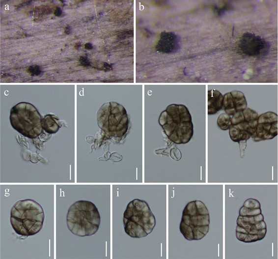
Saprobic on decaying wood. Sexual morph Undetermined. Asexual morph Colonies on natural substrate effuse, scattered, punctiform. Mycelium partly immersed, partly superficial, composed of brown, septate, branched hyphae. Conidiophores micronematous to semi-macronematous, mononematous, septate, branched, flexuous, subhyaline to pale brown. Conidiogenous cells monoblastic, integrated, terminal, subhyaline to pale brown, determinate, doliform. Conidia 17–33 × 13–24 μm (x̄ = 23.9 × 18.6 μm, n = 30), solitary, median brown, irregularly globose or subglobose, trapeziform, muriform, constricted at the septa, smooth-walled. Culture characteristics: Conidia germinating on water agar within 48 h. Germ tubes produced from the base of conidia. Mycelia superficial, circular, with entire edge, sparse, white to greyish white from above, grey from below. Material examined: CHINA, Guizhou Province, Dushan, on decaying wood in a bank of a small freshwater, 6 July 2018, N.G. Liu, DS020 (MFLU 19-2855, holotype), ex-type living culture, GZCC 20-0009.
Genbank numbers: ITS = MT002306, LSU = MN897833, SSU = MN901116, TEF1-α = MT023010.
Notes: Based on a blast search in NCBI, the closest hit using the LSU sequence are Conlarium duplicias- cosporum F. Liu & L. Cai (GenBank no. JN936993; Identities = 774/816 (95%), Gaps 2/816). Conlarium duplumascosporum was introduced by Liu et al. (2012) and its asexual morph was produced in culture. Morphologically, C. duplumascosporum and Pseudoconlarium punctiforme share similar conidial size (15.5–35 × 11–26.5 versus 17–33 × 13–24 μm), but P. punctiforme has more conidial septa than those of Conlarium duplumascosporum.

Fig. 121 Pseudoconlarium punctiforme (MFLU 19-2855, holotype). a, b Colonies in natural substrates. c–f Conidiophores, conidiogenous cells and conidia. g–k conidia. Scale bars: c–k = 10 μm
