Pseudocamarosporium corni Wijayaw., Camporesi & K.D. Hyde, in Wijayawardene, Hyde, Bhat, Camporesi, Schumacher, Chethana, Wikee, Bahkali & Wang, Cryptog. Mycol. 35(2): 187 (2014)
Index Fungorum Number: IF 550557; Facesoffungi number: FoF 0008
Holotype – MFLU 14-0089
Etymology – Named after the generic name of host, Cornus.
Saprobic on dead branch of Cornus sanguinea. Sexual state: Not observed. Asexual state: Conidiomata pycnidial, 180-270 µm diam., 190-250 µm high, gregarious, dark brown, immersed, unilocular, with centrally located papillate ostiole. Pycnidial wall multilayered, outer layer with 1-3 brown walled cells of textura angularis, inner wall with 1-4 layers, hyaline. Conidiophores reduced to conidiogenous cells. Conidiogenous cells enteroblastic, phialidic, percurrently proliferating, smooth, short, hyaline to pale brown. Conidia 11-16 × 6-8 µm (› = 13.4 × 7.2 µm, n = 20), oblong to ellipsoidal, mostly straight, rarely slight curved, muriform, with 1-3 transverse septa, with 1-2 longitudinal septa, occasionally widest at the middle, brown, smooth-walled.
Culture characteristics – on PDA white from above and very light brown from reverse, with thin mycelium, flat, attaining a diameter of 2.5 cm in 7 days at 18°C.
Material examined – Italy, Arezzo Province, Porrena-Poppi, on a dead branch of Cornus sanguinea L. (Cornaceae), 10 March 2013, E. Camporesi, NNW IT 1108 (MFLU 14-0089, holotype; isotype PDD 104440), ex-type living culture = MFLUCC 13-0541 = ICMP 20369 = GUCC 0010.
Notes – Hüseyinov (1968) reported Camarosporium corni-maris Hüseyın from Cornus sanguinea, which has larger conidia (40-64 × 16-23.5 µm). Our collection has smaller conidia, and molecular data analyses (Figs 2 & 3) show that it clusters in Montagnulaceae and not Camarosporium sensu stricto (Fig. 1). Hence, we introduce it as a new species in Pseudocamarosporium.
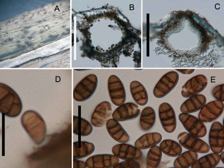
Fig. 1. Pseudocamarosporium corni (holotype). A. Conidiomata on Cornus sanguinea. B, C. Cross section of pycnidia. D. Developing conidium attach to conidiogenous cell. E. Conidia. Scale bar: B, C. 200 µm; D, E. 15 µm.