Podosphaera yulii S.-Y. Liu & P.-L. Qiu, sp. nov.
Index Fungorum number: IF 556431; MycoBank number: MB 556431; Facesoffungi number: FoF 06104; Fig. 36
Etymology: Refers to the name of Prof. Yu Li.
Holotype: HMJAU91796.
Mycelium amphigenous, effuse, persistent, often causing yellowish or dark brown discoloration and malformation of the hosts leaves. Hyphal appressoria indistinct nipple-shaped, solitary. Conidiophores arising from the upper surface of hyphal mother cells, (45–)50–105 × 9–12 μm and producing the conidia in chains with clear fibrosin bodies. Foot cells, cylindrical, 22–51(–56) × 7.8–12.7 μm, sometimes slightly swollen at the very base and the basal septum occasionally raising above the junction with the mother cell. Conidia ellipsoid or oval, with distinct fibrosin bodies, 18.6–29.1(–36.5) × 14–21 μm with a length/width ratio of mostly 1.2–2.1. Germ tubes of conidial germination, clavate or somewhat forked, straight or most curved, producing in single, opposite or double at the one end or both ends of the conidia. Chasmothecia 69–93 μm diam, amphigenous, gregarious, including numerous yellowish droplets, and containing one ascus each. Peridium cells of chasmothecia, irregularly polygonal. Appendages, 6–22 per chasmothecium, (41.7–)50–108(–148.4) μm, dark brown at the base, becoming paler towards the apex, apices dichotomously branched, 0.5–1.7 times the chasmothecial diam. Asci oval to subglobose, 49.3–75.5 × 46.5–65.2 μm, sessile or with a very short stalk, terminal oculus 5.5–17(–24) μm wide. Ascospores 8 per ascus, curved, crescent-shaped, very few ellipsoid, 18–25 × 9.2–13.2 μm, colorless, and emerging yellowish oil drops after heating in lactic acid.
Material examined: CHINA, Jilin Province, Changchun, Jilin Agricultural University, on Crataegus pinnatifida, 1 June 2018, P.L. Qiu (HMJAU91796, holotype).
Additional specimens examined: On Crataegus pinnatifida – CHINA, Heilongjiang, Heihe City, 1 September 2017, F.Y. Zhao & V. Nguyen HMJAU91798, HMJAU91799; Jilin Province, Changchun City, Jilin Agricultural University, 5 July 2018, P.L. Qiu (HMJAU91797).
GenBank numbers: ITS: MK409979, MK409981, MK409982, MK409980; LSU: MK734073, MK734075, MK734076, MK734074; IGS: MK955881.
Notes: Maximum parsimony phylogenetic analysis based on the ITS sequences demonstrated that Podosphaera yulii formed a single clade with 100% bootstrap support that is distant from P. clandestina, P. curvispora and P. paracurvispora (Fig. 37). The molecular analysis of 28S rDNA sequences suggested the four species clustered with each other but without bootstrap support (Fig. 38). The morphological characteristics and molecular analysis support that the powdery mildew on C. pinnatifida in this study is a new and undescribed species. P. yulii is clearly different from P. clandestina by having mostly curved ascospores, but a few straight, short foot-cells (up to 56 μm in P. yulii vs 35–100(–125) μm in P. clandestina) and more appendages (6–22 per chasmothecium in P. yulii vs 5–15 in P. clandestina). P. paracurvispora and P. curvispora also have curved ascospores. Tang et al. (2017) recorded that P. paracurvispora has foot-cells (13–)20–27(–32) μm in length which is shorter than P. yulii with foot-cells 22–51(–56) μm, and pale brown, curved, crescent-shaped ascospores that differ from P. yulii having curved and colorless ascospores. The conidial germination type (Fibroidium-type, singly formed subapically or laterally) within P. paracurvispora is also different from P. yulii. No specific anamorphic characters of P. curvispora were described in Braun and Cook (2012). P. yulii differs from P. curvispora by having smaller chasmothecia (69–93 μm diam vs (70–)100–150(–170) μm, Braun and Cook 2012).
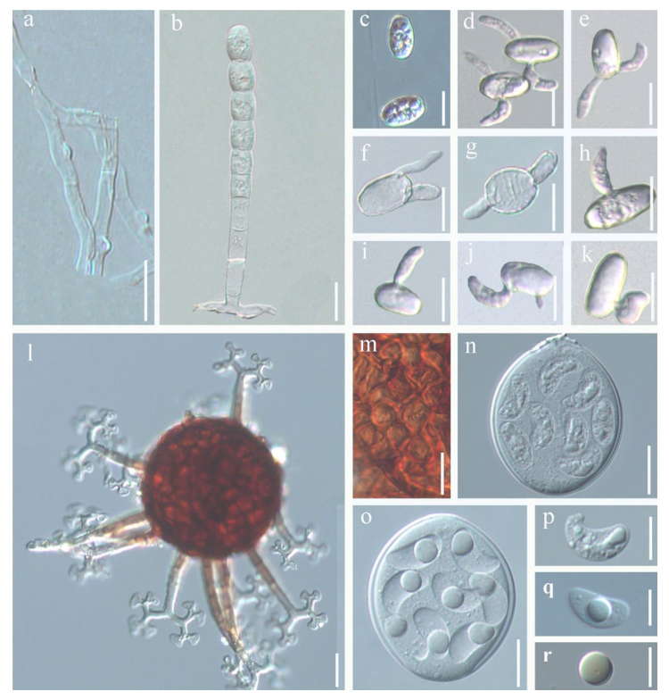
Fig. 36 Podosphaera yulii(HMJAU91796, holotype) on Crataegus pinnatifida. a Nipple-shaped hyphal appressoria. b Conidiophore. c Elliptical conidia. d–k Germ tubes of conidial germination. l Chasmothecia. m Peridium cells. n Ascus with 8 curved ascospores. o Ascus after heating. p Ascospore curved and colorless. q Ascospore with yellowish droplet after heating. r Droplet. Scale bars: a–o = 20
μm, p–r = 10 μm
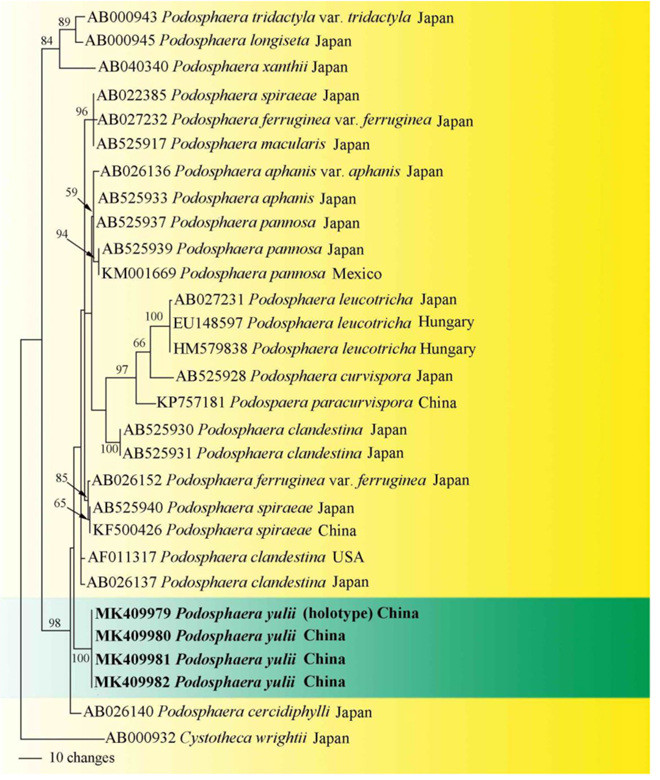
Fig. 37 Phylogram generated from maximum parsimony analysis based on ITS sequences of Podosphaera. Cystotheca wrightii (AB000932) was used as outgroup. Bootstrap values for maximum parsimony ≥ 50% were shown on the respective branches. The sequences of new taxon are shown in boldface
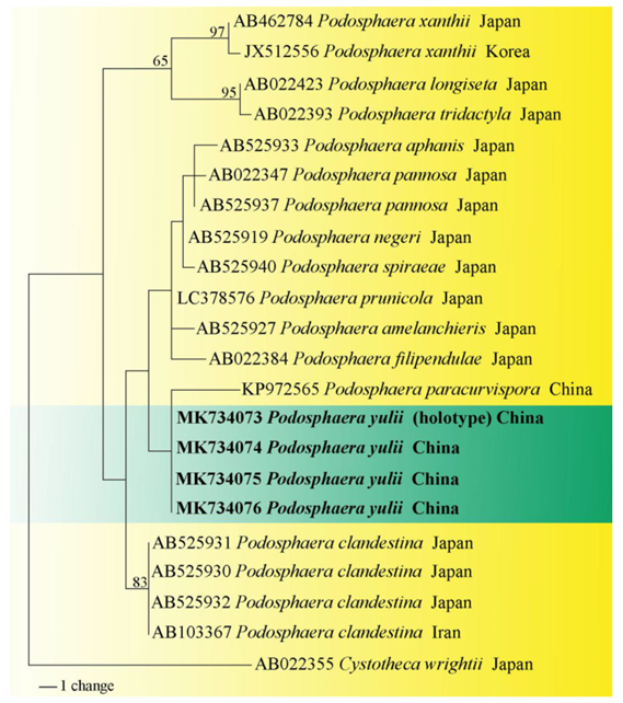
Fig. 38 Phylogram generated from maximum parsimony analysis based on LSU sequences of Podosphaera. Cystotheca wrightii (AB022355) was used as outgroup. Bootstrap values for maximum parsimony ≥ 50% were shown on the respective branches. The sequences of new taxon are shown in boldface