Phoma medicaginis Malbr. & Roum., Fungi Selecti Galliaei Exs. 37: no. 3675 (1886)
Index Fungorum number: IF 169294, Facesoffungi number: FoF00423; Fig. 1
Saprobic on Scabiosa sp., forming numerous, conspicuous, rounded to oval, dark brown, conidomata. Sexual morph Undetermined. Asexual morph coelomycetous. Conidiomata 100–150μm high, 150–250μm diam., pycnidial, solitary, separate, scattered or gregarious, globose, dark brown, immersed to semi-immersed, unilocular, thin-walled, with a single, papillate, centrally located ostiole. Peridium composed of 3–4 wall-layers, 15–30μm wide, with outer 1–2-layers dark brown, inner 1–2-layers hyaline, with thin walled cells textura angularis. Conidiophores reduced to conidiogenous cells arising from the innermost wall-layer cells of the conidiomata. Conidiogenous cells 4–7μm long×5–8μm wide, enteroblastic, phialidic, determinate, doliiform and hyaline. Conidia 10–20×3.5–5.5μm wide (x=15.5×5, n=20), ellipsoidal to ovoid, hyaline, straight or slightly curved, 1-septate, constricted at the septum, obtuse at both ends, thin-walled, smooth-walled, guttulate.
Culture characters: Colonies on PDA slow growing, reaching 25 mm diam. after one month at 25–30 °C, circular, grey to black, white at the edge, flattened with dense, filamentous, aerial, fluffy hyphae; reverse black in the middle, white at the edge, without any diffusible pigments.
Material examined: ITALY, Province of Ravenna [RA], Zattaglia, on dead twig of Scabiosa sp. (Caprifoliaceae), 30 December 2012, E. Camporesi IT-988 (MFLU 14–0812), living culture,MFLUCC 13–0485, ICMP 20794. GenBank ITS: KP711359; LSU: KP711364; SSU: KP711369; ibid. (KUN! HKAS 83971).
Notes: Phoma medicaginis was introduced by Malbranche & Roumeguère (1886). This species is cosmopolitan with a worldwide distribution and occurs on various hosts such as Brassica oleracea L., Medicago sativa Lam., Cicer arietinum L., Glycine soja sensu auct., Lathyrus odoratus L. and Medicago sativa L. (Sutton 1980; Aveskamp et al. 2010). Phoma medicaginis is an economically important pathogen, and most likely a species complex comprising morphologically indistinguishable, but genetically and biologically isolated species (Ellwood et al. 2006; Aveskamp et al. 2008, 2010). In addition, it has been described as an endophyte in the roots of Taxus globosa (Rivera-Orduña et al. 2011). The species is characterized by globose, large, pycnidia, with straight or slightly irregular, cylindrical conidia, often becoming 1-septate and variably guttulate (Sutton 1980). Our isolate (MFLUCC 13–0485) is morphologically similar with Ph. medicaginis, and the only distinguish morphological character is the dimension of conidia, but it should be noted that this character has proven not be very reliable in coelomycetes (Verkley et al. 2014). Phylogenetic analyses based on the combination of LSU and ITS sequence data coupled with morphological characters showed that the strain (MFLUCC13–0485) is conspecific with Phoma medicaginis. Hence, we provided here a description of this species for further reference.
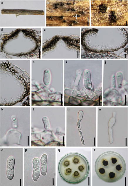
Fig. 1 Phoma medicaginis (MFLU 14–0812) a Specimen. b, c Black conidiomata on the host surface. d Vertical section of conidioma. e Ostiole. f, g Section of peridium. h–l. Conidiogenous cells and developing conidia. m, n Germinated spore. o, p Conidia. q, r Culture on PDA (note r; reverse). Scale bars: b=500μm, c=100μm, d=50μm, e–g=20μm, h–k=5μm, m–n=10μm, o–p=5μm, q–r=25 mm.