Phallus haitangensis H.L. Li, P.E. Mortimer, J.C. Xu & K.D. Hyde, sp. nov., Index Fungorum number: IF 552109
Etymology: The species epithet “haitangensis” refers to the place where the type species was collected.
Holotype: CHINA. Yunnan Province: Baoshan, Haitangwa Village, 25.266 °N, 99.300 °E, alt. 2473m, 8 April 2013, Huili Li (HKAS 88193).
Diagnosis: Receptacle strongly reticulate, golden orange, with a prominent opening at the apex. Pseudostipe creamy white, with a well-developed and light orange indusium, and a sac-like volva with rhizomorphs.
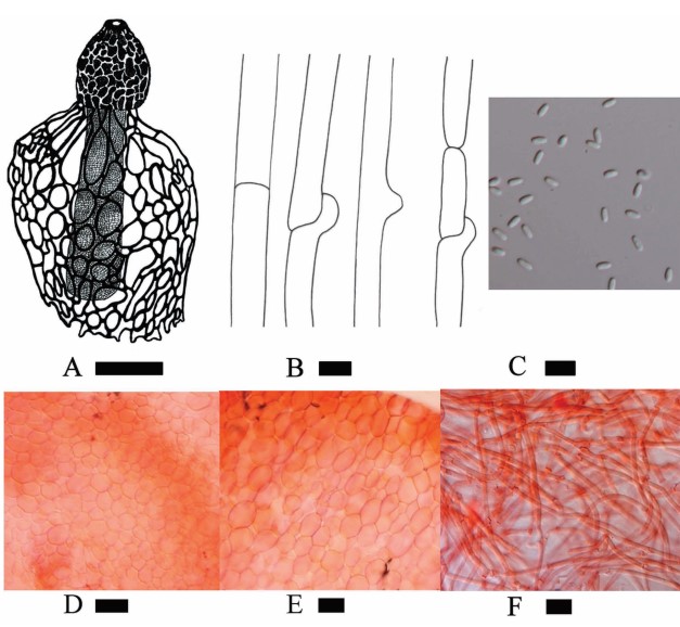
Sexual morph: Immature fruiting body not seen. Expanded basidioma up to 130–165 mm high, unbranched, with an indusium flaring out from beneath the receptacle, a stipe-like pillar and a volva with thin rhizomorphs. Receptacle 30–35 mm high × 25–50 mm wide, bell shape to umbrella shape, dark orange (5B7) to golden yellow (5B7), strongly reticulated, gold yellow (5B7) ridges, yellowish brown (5E5–7) sticky gleba in the pits, with a prominent hole at the apex with 1–8 mm diameter. Receptacle margin is lightly upturned. Pseudostipe creamy white (4A1), 100–132 mm high × 8–11/15–20/18–22 mm wide (apex-middle-base), nearly equal or lightly tapering upwards, fragile or soft, spongiform, holes in pseudostipe is 1–10 mm diameter. Indusium 85–95 mm long, 80–155 mm broad, pale orange (5A3) to light orange (5A4–5), well-developed, at first contracted under the edge of the receptacle, later expanding almost touching the ground. The holes of the indusium are nearly round to polygonal, 4–35 mm diameter and 1–4 mm thick, smooth edges. Volva present at base of pseudostipe, 30–140 mm high and 20–45 mm thick, yellowish white (4A2), smooth surface, attached to the substrate by a pinkish white (13A2) rhizomorph. Odor foetid. Basidiospores [100/4/4] 2.8–4.2 (3.4±0.3) µm length, 1.1–2.6 (1.7±0.3) µm width, L=3.4 µm, w=1.7 µm, Q=2, elongate or cylindrical, hyaline, smooth surface. Cells of pseudostipe 15–38.8 (28±6.2) µm diameter, bubble-like or foam-like, hyaline, smooth surface. Cells of indusium 21.2–41.6 (30.1±5.7) µm diameter, bubble-like or foam-like, hyaline, smooth surface. Hyphae of volva 1.8–3.8 (2.6±0.5) µm width, tubular and branched, septate with clampconnection, hyaline, smooth surface (Fig.3).
Habitat and Distribution: solitary or scattered on soil with decaying litter under P. armandii trees, Yunnan Province, China.
Material examined:—CHINA. Yunnan Province: Baoshan, Haitangwa Village, 25.266°N, 99.300°E, alt. 2473m, 8 April 2013, huili Li (hKAS 88193, holotype); Ibid., Haitang Village, 4 August 2013, huili Li (hKAS 88191, paratype); Ibid. 26 August 2014, huili Li (hKAS 88197, paratype); Ibid. 2 September 2014, huili Li (hKAS 88199, paratype).
Notes: The main distinguishing characteristics of Phallus haitangensis are the golden orange receptacle that is strongly reticulate, with a somewhat prominent apex, which has a light orange opening, and a light orange indusium with rounded to polygonal meshes without serrated margins (Fig. 2), and a creamy white spongy pseudostipe which tapers upwards. The results of the phylogenetic tree showed that P. haitangensis grouped with P. serrata, P. mengsongensis and P. cinnabarinus, however P. serrata has a white and half-egg receptacle, a white indusium with serrated margins of the holes, and larger basidiospores (Li et al. 2014); P. mengsongensis has small fruiting bodies without an indusium (Li et al. 2014); the receptacle of P. cinnabarinus is cinnabar-red and the pseudostipe is pale cinnabar-red and shorter than that in P. haitangensis, and the indusium of P. cinnabarinus is also cinnabar-red above to paler below, and the basidiospores of P. cinnabarinus are brownish green (Lee 1957, Hemmes & Desjardin 2009).
FIG 2. Plate of the Phallus haitangensis (HKAS 88193). A Mature fruiting body of P. haitangensis. B Hole at the apex of the receptacle. C Indusium. D Receptacle. E Pseudostipe. F Volva and rhizomorph. Photos by Huili Li.
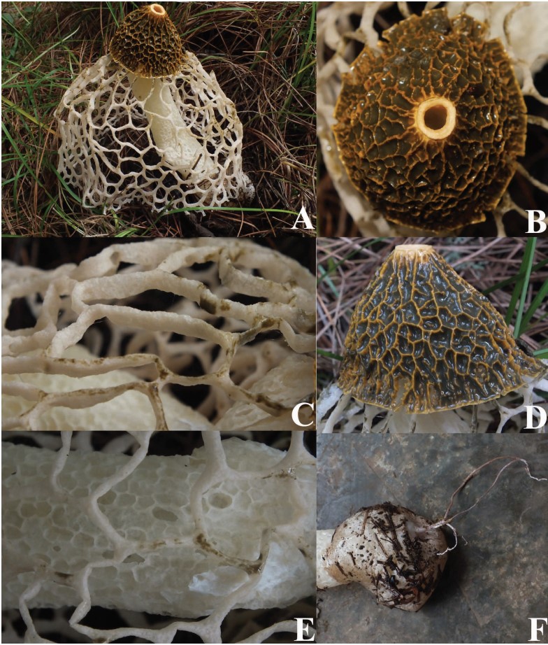
FIG 3. Morphological characteristics of Phallus haitangensis. A Line drawing of basidiocarp. B Line drawing of hyphae of volva. C Basidiospores. D Cells of pseudostipe. E Cells of indusium. F Hyphae of volva. Scale bars: A=20 mm, B=20 µm, C=5 µm, D=40 µm, E=40 µm, F=10 µm. Tissues of the structures D, E and F were mounted in Congo Red for microscopic observation. Drawings by Samantha C. Karunarathna and photos by Huili Li.