Phaeoisaria goiasensis H.M. Silva, A.D. Cavalcanti & J.D.P. Bezerra, sp. nov., MycoBank
Number: MB840294
Description
Isolated from a Petri dish with culture medium storage in a fridge. Asexual morph: Hyphae
hyaline to pale brown with age, smooth wall, septate, 1.5–2.5(–3) µm wide. Synnemata erect,
brown, smooth, indeterminate, composed of compactly and parallels conidiophores and
commonly with conidiogenous cells in the above half, 93–147 × 3.5–4.5 µm. Conidiophores
straight or slightly curved, septate, reduced to conidiogenous cell, cylindrical, hyaline to pale
brown, smooth wall, (10–)15–49(–72) × (1.5–)2–2.5(–3.5) µm. Conidiogenous cells polyblastic,
integrated, terminal or intercalary, cylindrical, hyaline, smooth wall, (3–)3.5–9.5(–15.5) ×
(1.5–)2–3(–3.5) µm, forming conidia on denticles, 1–2 µm long, 0.5–1 µm wide, scattered or
clustered in the apical region. Conidia ellipsoidal to obovoid, straight or slightly curved, rounded
at the ends or occasionally tapering toward the base, hyaline, aseptate, guttulate, smooth wall,
(4.5–)7.5–9(–10.5) × (2–)2.5–3(–4) µm. Chlamydospores terminal, globose, pyriform, first
hyaline and becoming brown to dark brown with age, (8–)8.5–10.5(–17) × (2–)7–8(–8.5) µm.
Sexual morph: Not observed.
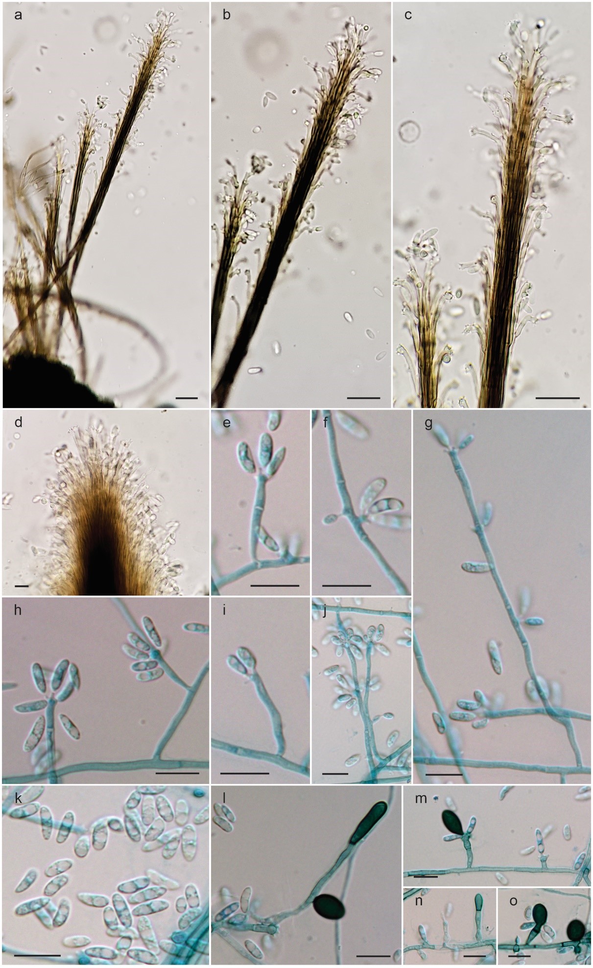
Fig. x. Phaeoisaria goiasensis (UFG 71083, holotype). A–C Synnemata. D Details of a synnema. E–J Conidiophores and conidia. K Conidia. L–O Chlamydospores and conidia. Scale bars: 10 μm.