Pestalotiopsis subshorea Yong Wang bis, Yu Song, K. Geng & K.D. Hyde, in Ariyawansa et al., Fungal Diversity: 10.1007/s13225-015-0346-5, [138] (2015)
Index Fungorum number: IF551449; Facesoffungi number: FoF01045; Fig. 1
Etymology – The specific epithet is referring to Pestalotiopsis shorea, which is similar to this new taxon.
Holotype – HGUP d4118 Endophyte in leaves of Micelia hedyosperma Law. Sexual morph: Undetermined. Asexual morph: Conidiophores indistinct. Conidiogenous cells discrete, simple, filiform, thinwalled, hyaline. Conidia fusoid, straight to slightly curved, 4- septate, 18–24.5×5.5–7 μm (x̄ = 22×6 μm), basal cell conic, hyaline or pale olivaceous, thin-walled and verruculose, 3.3– 5.3 μm long (x̄ = 4.5 μm), with three median cells, doliform to cylindrical, constricted at the septa, concolourous, olivaceous to brown, septa and periclinal walls darker than the rest of the cell, wall rugose, together 12–16 μm long (x̄ = 14 μm) second
cell from base 3.5–6 μm (x̄ = 5 μm); third cell 4–6 μm (x̄ = 4.5 μm); fourth cell 4–6 μm (x̄ =5 μm); apical cell hyaline, conic to subcylindrical 3–5.3 μm long (x̄ = 4 μm); with 2–3 tubular apical appendages, arising from the apex of the apical cell, 10–28 μm long (x̄ = 17 μm), unequal; basal appendage present 2–5.5 μm (x̄ = 3.5 μm), rarely absent.
Culture characteristics: Colonies on PDA reaching 7 cm diam. after 6 days at 25 °C, edge entire, whitish, with dense aerial mycelium on surface, fruiting bodies black; reverse of culture whitish.
Material examined – CHINA, Guizhou Province, Guiyang City, Xiaohe District, on living leaves of unidentified tree, Kun Geng, 25 September 2012 (HGUP d4118, holotype); ex-type culture, HGUP4118; CHINA, Guangxi Province, on living leaves of Micelia hedyosperma (Magnoliaceae), Jiguang Wei, 2011 (HGUP4279, paratype).
Notes – Phylogenetic analysis of three combined gene loci (ITS+β-tubulin+EF) show Pestalotiopsis subshorea (HGUP 4118 and HGUP 4279) forming a separate branch as a sister taxon to P. shorea (Fig. 2). Pestalotiopsis shorea was isolated from Shorea obtusa in Thailand (Song et al. 2014). Both species produce concolourous conidia and their conidial size overlap. The morphological distinction of P. subshorea and P. shorea is based on the length of apical and basal appendages. Apical appendages of P. subshorea are longer than P. shorea (3–5 μm long), but the basal appendage is shorter than P. shorea or lacking. Thus, we confirm the novelty of P. subshorea.
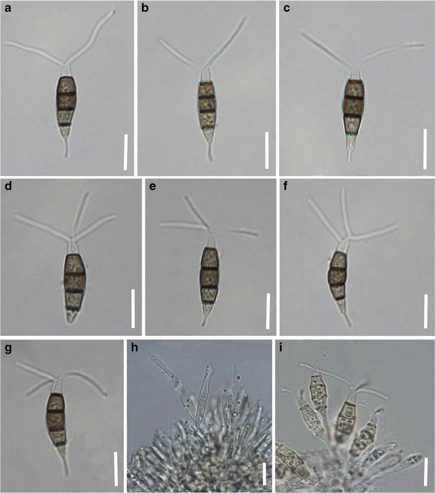
Fig. 1 Pestalotiopsis subshorea (holotype) a–g Conidia h, i Conidiophores / conidiogenous cells. Scale bars=10 μm
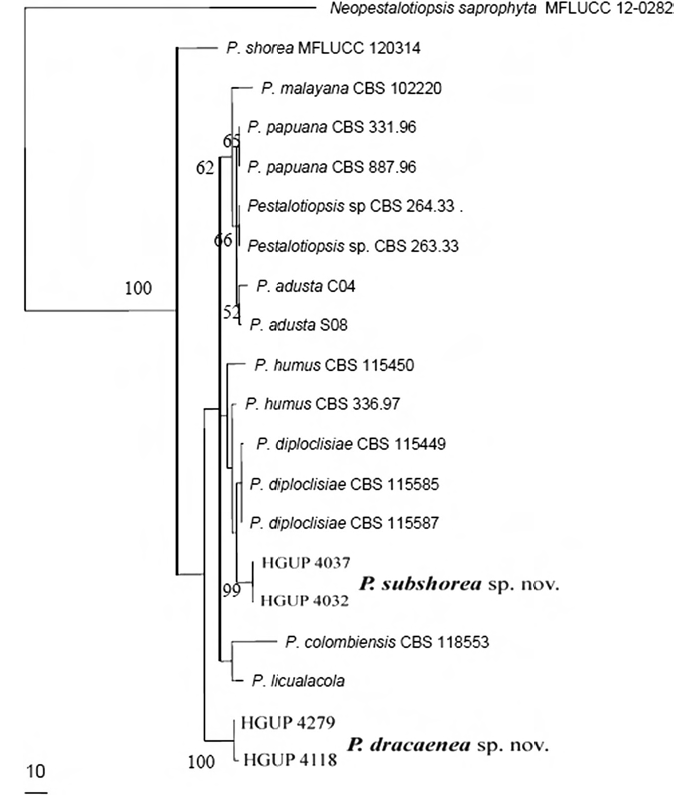
Fig. 2 Phylogenetic tree for Pestalotiopsis based on combined sequences of ITS, β-tubulin and EF by Maximum Likelihood (ML) method on MEGA 6.0. Bootstrap support values <50 % were not shown. The tree is rooted with Neopestalotiopsis saprophyta (MFLUCC 12-0282)