Pestalotiopsis rhodomyrtus Yu Song, K. Geng, K.D. Hyde & Yong Wang bis [as ‘rhodomyrtus‘], in Song, Geng, Zhang, Hyde, Zhao, Wei, Kang & Wang, Phytotaxa 126(1): 27 (2013)
Index Fungorum number: IF 804968; MycoBank number: MB 804968; Facesoffungi number: FoF 09398;
Etymology — In reference to the host, Rhodomyrtus tomentosa, from which this fungus was first isolated
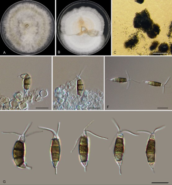
Pathogenic to host leaves. Asexual state: Conidiophores indistinct. Conidiogenous cells discrete, hyaline, simple, filiform. Conidia 19.7–26.3 × 4.9–6.7μm (n = 30, x̄ = 23.00 × 5.76 μm), fusoid, straight to slightly curved, 4-septate; basal cell conic to obconic, hyaline or pale brown, smooth, thin-walled, 3.6–6.4 μm long (n = 30, x̄ = 4.79 μm); three median cells 12.9–16.8 μm long (n = 30, x̄ = 15.04 μm), brown, septa and periclinal walls darker than the rest of the cell, concolorous, verruculous; second cell from base 4.3–5.8 μm long (n = 30, x̄ = 5.12 μm); third cell from base3.5–5.7 μm long (n = 30, x̄ = 4.73 μm); fourth cell from base 4.3–5.6 μm long (n = 30, x̄ =4.88 μm); apical cellhyaline, obconic to subcylindrical, 2.7–4.1 μm long (n = 30, x̄ = 3.56 μm); with 2–3 tubular appendages, arising from the apex of the apical cell, 7.5–14.9 μm long (n = 30, x̄ = 10.54 μm), unequal; one basal appendage present, 2.8–4.9 μm long (n = 30, x̄ = 3.65 μm), filiform. Sexual morph unknown.
Culture characters — Colonies on PDA reaching 7 cm diam. after 8 days at 25°C, with edge crenate, whitish, aerial mycelia on surface, fruiting bodies black, gregarious; reverse of colony pale orange.
Material examined — China, Guizhou Province, Zunyi City, Suiyang County, Kuankuoshui Natural Reserve, on diseased leaves of Cyclobalanopsis augustinii (Fagaceae), 23 November 2019, Dan-ran Bian (culture CFCC 55052); ibid., Shaanxi Province, Hanzhong City, Foping County, Dongshan Mountain, on diseased leaves of Quercus aliena (Fagaceae), 7 September 2019, Yong Li (culture CFCC 54733).
Distribution — China
Sequence data — ITS: OM746310 (ITS1/ITS4); tef1: OM840082 (EF1-728F/EF2); tub2: OM839983 (Bt2a/Bt2b)
Notes — Conidia of P. rhodomyrtus are similarin shape to those of P. trachicarpicola, P. rosea and P. adusta. However, the conidia of P. rhodomyrtus are bigger than those of P. rosea and P. adusta. The apical appendages of P. rhodomyrtus are shorter thanthose of P. trachicarpicola and P. rosea. Pestalotiopsis rhodomyrtus possess only one basal appendage, but P. trachicarpicola rarely has two, and the length of their basal appendage is different.

Fig. 19. Morphology of Pestalotiopsis rhodomyrtus (CFCC 55052). A. Colony on PDA after 10 d at 25 °C; B. Colony on MEA after 10 d at 25 °C; C. Conidiomata formed on PDA; D, E. conidiogenous cells giving rise to conidia; F, G. conidia. — Scale bars: C = 300 μm; D–G = 10 μm.
