Pestalotiopsis montellica (Sacc. & Voglino) Tak. Kobay., Trans. Mycol. Soc. Japan 15(4): 381 (1974)
Index Fungorum number: IF319429 ; Facesoffungi number: FoF01043; Fig. 1
= Pestalotia montellica Sacc. & Voglino, Atti Accad. Sci. Soc. Ven.-Trent. Sci. Nat. 9(2): 9 (1892)
Saprobic on dead plant material. Sexual morph: Undetermined. Asexual morph: Conidiomata pycnidial in culture on PDA, globose, scattered and confluent, semi-immersed, dark brown. Conidiophores indistinct. Conidiogenous cells lageniform to subcylindrical, colourless, smooth, proliferation once or twice. Conidia fusoid to ellipsoid, straight to slightly curved, 4-septate, 23–29×5.5–7 μm (x̄ = 25×6.1 μm), basal cell obconic, colourless, thin- and verruculose, 5–6 μm long (x̄ = 5.3 μm), with three median cells, subcylindrical, with thick verruculose walls, constricted at the septa, concolourous, pale brown, together 14–16.5 μm long (x̄ = 15.2 μm) second cell from base 4.5–6.5 μm(x̄ = 5.8 μm); third cell 4–6 μm(x̄ = 5.2 μm); fourth cell 5–6μm (x̄ = 5.2 μm); apical cell colourless, obconic, acute at the apex, 4–5 μm long (x̄ = 4.3 μm); with 4 tubular appendages, 14–20 μm long, one arising from the apex and rest arising from just above the
septum separating upper median and apical cell; basal appendage present, 4–5 μm long (x̄ = 4.5 μm).
Cultural characteristics: Colonies on PDA reaching 7 cm diam. after 7 days at 25 °C, edge lobate, whitish yellow, with dense, aerial mycelium on surface, with black, gregarious fruiting bodies; reverse of the colony whitish yellow.
Material examined – CHINA, Yunnan Province, on dead plant material,WenpingWu (NN042849, MFLUCC 12-0279).
Notes – Pestalotiopsis montellica was described from the leaves of Quercus sp. in Italy and is well characterized and easily recognizable by the unique appendages attached to the apical cell. The arrangement of apical appendages in P. montellica is comparable with P. jesteri (Strobel et al. 2000). However, P. jesteri Strobel et al. differs from P. montellica by the presence of knobbed apical appendages. A megablast search of the NCBIs GenBank nucleotide sequence
database using the sequences of P. montellica retrieves as closest hits P. jesteri (ITS: GenBank KM199380; Identities= 512/536(96 %), Gaps=8/536(1 %); TUB GenBank KM199468; Identities=403/444(91 %), Gaps=7/444(1 %)). The attachment and arrangement of apical appendages at the apical cell are noticeably distinct in both species. Previously Maharachchikumbura et al. (2012) misidentified this isolate as a P. montellica.
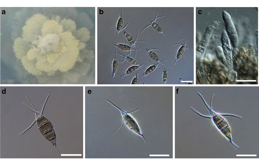
Fig. 1 Pestalotiopsis montellica (MFLUCC 12–0279) a Colony on PDA b Conidia on PDA c Conidiogenous cells d–f Conidia. Scale bars: b–f= 10 μm