Periamphispora phacelodes J.C. Krug, Myco- logia 81(3): 476 (1989)
MycoBank number: MB 135949; Index Fungorum number: IF 135949, Facesoffungi number: FoF 10044; Fig. 61
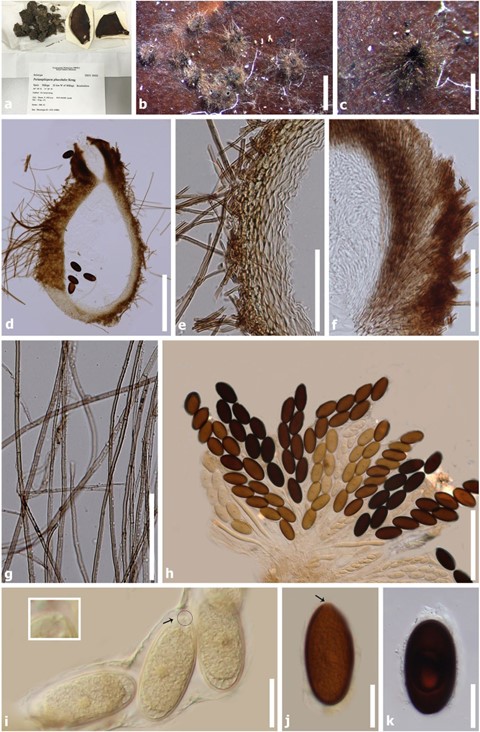
Coprophilous. Sexual morph : Ascomata 650–700 × 360–380 µm (x̄ = 670 × 370 µm, n = 5), perithecial, solitary to scattered, superficial to semi-immersed, globose to subglobose, brown, membranaceous to coriaceous, tuberculate, ostiolate, with necks. Necks brown to dark brown, surrounded by numerous, pale brown, filiform, septate, hairs 1.5–3.5 µm wide. Peridium 33–55 µm (x̄= 43 µm, n = 30) wide, outer layer composed of pale brown to reddish brown cells of textura porrecta to textura angularis; inner layer composed of hyaline cells of textura prismatica. Paraphyses numerous, filiform, septate, evanescent. Asci 200–300 × 40–65 µm (x̄ = 250 × 55 µm, n = 30) ascospore-bearing part, 8-spored, unitunicate, cylindrical to clavate, pedicellate, about 100 µm long, apex rounded. Ascospores (30–)40–45(–50) × 20–25(–30) µm (x̄ = 42 × 23 µm, n = 50),bi-seriate, ellipsoidal to oval, aseptate, ends rounded, with a germ pore at the apex, hyaline when young, becoming brown to reddish brown, rough-walled, surrounded by hyaline, gelatinous sheath, 2–5 µm wide. Asexual morph: Undetermined.
Material examined – Spain, Málaga, about 20 km W of Málaga, Benalmádena (36°36’N, 4°34’W), on horse dung, 30 April 1971, P. Blaser (TRTC-55023, holotype).
Known hosts and distribution – On horse dung in Spain (type locality) (Krug 1989).
Notes – We re-examined the type, Periamphispora phacelodes 55023, which was grown on dung extract agar medium culture (Weitzman and Silva-Hutner 1967), and it is well preserved.

Fig. 61 Periamphispora phacelodes: (TRTC-55023, holotype). a Herbarium material label. b Ascomata on Weitzman and Silva-Hutner’s agar medium. c Appearance of ascoma with hyphae. d Ascoma in cross section. e Peridium. f Perithecial neck with periphyses. g Hyphae. h
Asci. i–k Ascospores with germ pore (arrowed and enlarged the apical germ pore). Scale bars: b = 1 mm, c = 500 µm, d = 200 µm, e–g = 50 µm, h = 100 µm, i–k = 20 µm
