Paraphaeosphaeria spartii W.J. Li, Camporesi & K.D.Hyde.
Index Fungorum number: IF550917, Facesoffungi number: FoF00421; Fig. 2
Etymology – Referring to the host, Spartium, on which the fungus was found.
Holotypus – MFLU 14–0810
Saprobic on dead stem of Spartium junceum L. Sexual morph Undetermined. Asexual morph coelomycetous. Conidiomata 200 – 300 (x̄ = 250) μm diam., 250 – 300 (x̄ = 260)μm high, pycnidial, epidermal to subepidermal, scattered, globose to subglobose, unilocular, pale brown, ostiolate, appearing pruinose, thick-walled. Ostiole circular, papillate. Peridium 10 – 20 μm wide, composed of 3 – 4 – layers, with outer 1 – 3 – layers brown and inner 1 – 2 – layers hyaline, thin-walled cells of textura angularis. Conidiophores reduced to conidiogenous cells, arising from basal layers of conidioma. Conidiogenous cells 2 – 6 × 3 – 6.5 (x̄ = 4 × 4.5) μm, enteroblastic, phialidic with an inconspicuous periclinal thickening, cylindrical to subcylindrical, or subcylindrical to ampulliform, integrated, hyaline, smooth-walled. Conidia 4 – 7.5 × 3.5 – 5 μm (x̄ = 6 × 4, n = 50), subglobose to ellipsoid or obovoid, hyaline when young, becoming pigmented to pale brown at maturity, smooth-walled, guttulate, aseptate, thinwalled.
Culture characters – Colonies on PDA slow growing, reaching 10 – 20 diam. after 3 weeks, glabrous and with colourless margin; mycelium immersed, initially colourless, later becoming yellowish aerial mycelium, non sporulating.
Material examined – ITALY, Province of Forlì-Cesena [FC], Santa Sofia, Collina di Pondo. on dead stem of Spartium junceum L. (Fabaceae), 16 October 2012, E. Camporesi IT-816 (MFLU 14–0810, holotype); ex-type living culture, MFLUCC 13–0398, ICMP 20789. GenBank ITS:KP711357; LSU: KP711362; SSU: KP711367; ibid. (KUN!HKAS 83969, isotype).
Notes – Morphologically, Paraph. spartii shares similarities with Paraph. sporulosa in having subglobose to ellipsoid or obovoid, aseptate conidia with one large and often also 1 – 2 additional smaller oil-droplets. Parac. spartii can be distinguished from Paraph. sporulosa by its conidiogenous cells. Parap. spartii has subcylindrical to ampulliform, integrated, phialidic conidiogenous cells, with an inconspicuous periclinal thickening and collarette. Paraph. sporulosa has globose to ampulliform, hyaline, discrete, conidiogenous cells with 1–2 percurrent proliferations. Phylogenetically, Paraph. spartii is also distinct from any other species within Paraphaeosphaeria (Fig. 1)
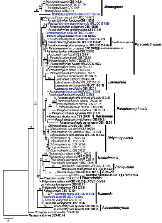
Fig. 1 Phylogram generated from Maximum likelihood analysis based on combined SSU, LSU, ß- tubulin and ITS sequence data of Didymosphaeriaceae. Maximum likelihood bootstrap support values greater than 50 % are indicated above and below the nodes, and branches with Bayesian posterior probabilities greater than 0.95 are given in bold. The ex-types (reference strains) are in bold; the new isolates are in blue. The tree is rooted with Massarina eburnea CBS 473.64.
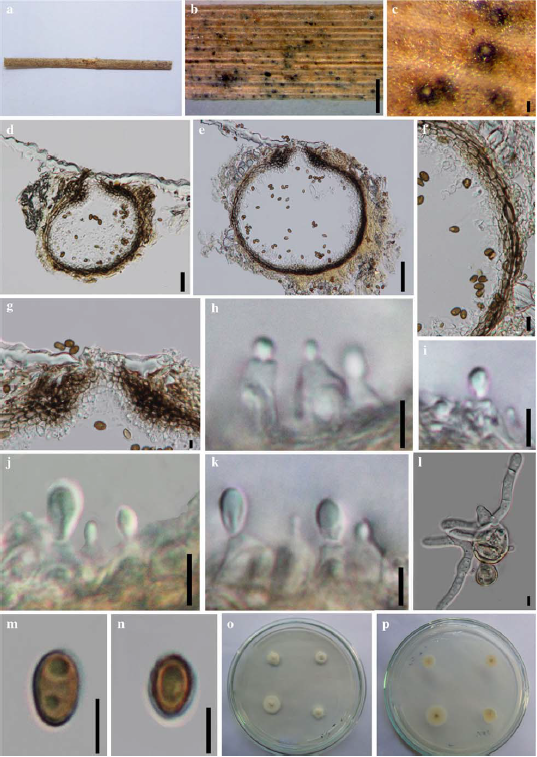
Fig. 2 Paraphaeosphaeria spartii (holotype). a Specimen b – c Black conidiomata on host surface d, e Vertical section of conidioma f Section of peridium g Ostiole h – k Conidiogenous cells and developing conidia l Germinated spore m – n Conidia o. p Culture on PDA. Scale bars: b = 500 μm, c = 200 μm, d = 100 μm, e = 5 μm, f = 5 μm, g = 10 μm, h – i = 5 μm, j – n = 5 μm.