Paraeutypella guizhouensis L.S. Dissan., J.C. Kang & K.D. Hyde, in Dissanayake, Wijayawardene, Dayarathne, Samarakoon, Di, Hyde & Kang, Biodiversity Data Journal 9: e63864, 12 (2021)
Index Fungorum number: IF 557953; MycoBank number: MB 557953; Facesoffungi number: FoF 09148;
Etymology– The specific epithet guizhouensis refers to the locality in which the fungus was collected.
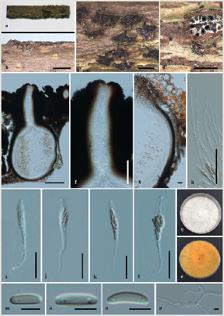
Saprobic on dead twigs. Sexual morph: Stromata immersed in bark of dead branches, erumpent, aggregated, circular to irregular, superficial, carbonaceous. Ascomata 590–600 × 470–480 μm (x̅ = 595 × 475 µm, n = 10), perithecial, with groups of 6–12 perithecia arranged in a valsoid configuration, subglobose, clustered, immersed in stromata, ostiolate. Neck 400–418 μm long (x̅ = 409 µm, n = 10), papillate, central ostiolar canal filled with periphyses, 3–4 sulcate. Peridium 22–35 μm wide, composed of two layers of textura angularis; inner layer cells light brown to hyaline, outer layers cells dark brown to black. Hamathecium hyaline. Paraphyses 1–2 μm wide (x̅ = 1.5 µm, n = 10), arising from base of perithecia, long, narrow, unbranched, septate, guttulate, narrowing and tapering towards apex. Asci 55–80 × 5– 9 μm (x̅ = 67.5 × 7 μm, n = 20), 8-spored, unitunicate, thin-walled, clavate to cylindrical clavate, long pedicellate (25–30 μm), with a J- apical ring. Ascospores 7–11 × 1–3 μm (x̅ = 9 × 2 μm, n = 30), overlapping biseriate, allantoid, hyaline to light brown, smooth, aseptate, usually with 2–3 guttules. Asexual morph: Undetermined.
Culture characteristics – Colonies on PDA, reaching 21 mm diam. after 2 weeks at 20– 25oC, medium dense, circular to slightly irregular, slightly raised, cottony surface; colony from above: at first white, becoming buff; from below: yellowish-white at margin, yellow to brown at centre; mycelium yellowish.
Material Examined – China, Guizhou Province, Guiyang, Guizhou University Garden (North), on dead twigs, L.S.Dissanayake, CLD018 (HMAS 290654, Holotype), ex-type KUMCC 20– 0016; ibid., (HKAS 290655, isotype), living culture KUMCC 20-0017.
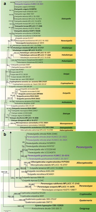
Notes – Paraeutypella guizhouensis resembles P. vitis, which comprises stromata that are erumpent through bark, with elongated perithecial necks and allantoid, slightly to moderately curved ascospores (Glawe and Jacobs 1987). However, P. guizhouensis differs from P. vitis in having comparatively longer ostiolar necks and longer asci (55– 80 × 5–9 μm), while P. vitis has comparatively shorter ostiolar necks and shorter asci (40–46 × 6–8 μm) (Glawe and Jacobs 1987). Paraeutypella vitis (UCD2428TX) differs phylogenetically from our new taxon in 14% (80/576) base pairs in the ITS and 10% (42/405) base pairs in β-tubulin. Thus, P. guizhouensis is introduced as a new species in Paraeutypella, based on its morphology, base pair differences and phylogenetic analyses (94% ML, Fig. 1).

Figure 1. Phylogram generated from Maximum Likelihood (RAxML) analysis, based on ITS- β-tubulin matrix. ML bootstrap supports (≥ 70%) and Bayesian posterior probability (≥ 0.95) are indicated as ML/BYPP. The tree is rooted to Kretzschmaria deusta (CBS 826.72) and Xylaria hypoxylon (CBS 122620). Newly-generated strains are in red and type strains are in bold. The asterisks represent unstable species.

Figure 3. Paraeutypella guizhouensis (HMAS 290654, holotype) a–c. stromata on substrate; d. cross section of a stromata; e. vertical section through an ascostroma showing ostioles and perithecia; f. ostiolar canal; g. peridium; h. paraphyses; i–l. asci; m–o. ascospores; p. germinating ascospore; q, r. cultures on PDA from above and below after 6 weeks. Scale bars: 500 µm (b–d), 200 µm (e), 100 µm (f), 20 µm (g–l), 5 µm (m–p).
