Paraconiothyrium nelloi W.J. Li, Camporesi & K.D.Hyde.
Index Fungorum number: IF550918, Facesoffungi number: FoF00422; Fig. 2
Etymology – Named after Camporesi Nello, who collected the sample from which the species was isolated.
Holotypus – MFLU 14–0813
Saprobic on dead stem of Spartium junceum L. Sexual morph Undetermined. Asexual morph coelomycetous. Conidiomata 250 – 350 μm high, 200 – 300 μm diam., pycnidial, solitary, immersed, globose to obpyriform, unilocular, centrally ostiolate, thick-walled. Peridium 15–25μm wide, 4–5- layered, composed of outer 3 – 4 – layers brown and inner 1 – 2 – layers hyaline, thin-walled cells of textura angularis. Conidiophores reduced to conidiogenous cells, arising from the base and sides of the conidioma. Conidiogenous cells 3.5 – 10 μm long × 5 –10 μm wide, enteroblastic, phialidic, determinate, ampulliform, lining the inner wall layer of the pycnidium, hyaline, smooth. Conidia 6.5 –8.5 × 5 – 6 μm (x̄ = 7.5 × 5.5; n = 10), globose to obovate, thick-walled, smooth-walled, onecelled, hyaline when young, becoming dark brown at maturity.
Culture characters – Culture on PDA slow growing, reaching 10 – 15 mm diam. after one week, circular, yellowish in the centre, with whitened edge, after one month, sparse, aerial, filamentous, no pigments produced.
Material examined – ITALY, Province of Forlì-Cesena [FC], Premilcuore, Fiumicello, on dead twig of Spartium junceum L. (Fabaceae), 13 January 2013, E. Camporesi IT-1008(MFLU 14–0813, holotype), ex-type living culture, MFLUCC 13–0487, ICMP 20796. GenBank ITS:KP711360; LSU: KP711365; SSU: KP711370; ibid. (KUN!HKAS 83972, isotype).
Notes – Paraconiothyrium was introduced by Verkley et al.(2004) to accommodate four species, namely Parac. estuarinum (type specie), Parac. Brasiliense, Parac. cyclothyrioides and Parac. fungicola. Subsequently, the genus was expanded to include four more species, viz. Parac. africanum, Parac. babiogorense, Parac. hawaiiense and Parac. variabile (Damm et al. 2008; Budziszewska et al. 2011). Presently, Paraconiothyrium comprises 21 species (de-Gruyter et al. 2013; Ariyawansa et al. 2014b; Verkley et al. 2014), including Parac. nelloi and the sexual species Parac. thysanolaenicola described in this paper. The morphological characters of Paraconiothyrium are variable. The conidiomata can be eustromatic to pycnidial, the conidiogenous cells are phialidic or annelidic, and the conidia smooth-walled or minutely warted and hyaline to brown at later stages of development (Verkley et al. 2004; Gruyter et al. 2013). The description of Parac. nelloi fits well with this generic concept, and Parac. nelloi shares similarities with Parac. fuckelii in having pycnidial conidiomata with a single ostiole, and subglobose to ellipsoid or obovoid conidia (Gruyter et al. 2013; Verkley et al. 2014). Parac. nelloi differs from Parac. fuckelii by the conidiogenous cells, which are phialidic in Parac. nelloi and annelidic in Parac. fuckelii. Combined phylogenetic analyses of ITS, SSU and LSU sequence data (Fig. 1) show that Parac. nelloi is distinct from any other species of Paraconiothyrium. Based on the morphological characters together with molecular sequence data, Parac. nelloi is introduced as a new species.
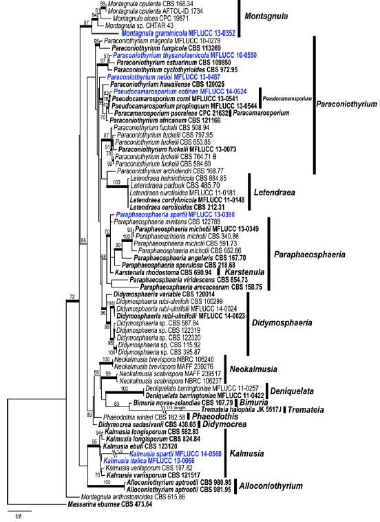
Fig. 1 Phylogram generated from Maximum likelihood analysis based on combined SSU, LSU, ß- tubulin and ITS sequence data of Didymosphaeriaceae. Maximum likelihood bootstrap support values greater than 50 % are indicated above and below the nodes, and branches with Bayesian posterior probabilities greater than 0.95 are given in bold. The ex-types (reference strains) are in bold; the new isolates are in blue. The tree is rooted with Massarina eburnea CBS 473.64.
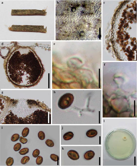
Fig. 2 Paraconiothyrium nelloi (holotype) a Specimen b Black conidiomata on the host surface c Section of peridium d Vertical section of conidiomata e, f Conidiogenous cells and developing conidia g Ostiole h Germinated spore i–k Conidia l Culture on PDA. Scale bars: b = 200 μm, c = 20 μm, e – f = 5 μm, g = 50 μm, i – k = 5 μm, l = 25 mm.