Otospora bareai sp. nov. J. Palenzuela, N. Ferrol & Oehl FIGS. 1–11
MycoBank number: MB 533135; Index Fungorum number: IF 533135; Facesoffungi number: FoF;
Sporocarpia ignota. Sacculus sporifer subhyalinus vel pallido-luteus vel luteus, globosus (150–210 mm diam) vel subglobosus (140–190 × 150–215 mm diam) et formationi sporae praecedens. Sporae singulae lateraliter formatae ad hypham in 30–90 mm distantia ad sacculum terminalem, flavae-brunneae vel fulvaebrunneae vel brunneae, globosae (150–200 mm diam) vel subglobosae (145–185 × 175– 210 mm diam) vel ovoideae vel ellipsoideae vel irregulares. Sporae non colorantes reagente Melzeri et tunicis duabus: tunica exterior et interior. Tunica exterior in totum 5– 12 mm crassa, stratis quartuoribus: stratum exterior hyalinum, tenue et evanescens; stratum secundum laminatum vel unitum, flavum vel fulvum (2.0–)3.0–4.5(–6.5) mm crassum, semi-persistens; stratum tertium laminatum, fulvum-brunneum vel brunneum et (2.5–)4.0–6.0 mm crassum et persistens; stratum interior (flavum vel) fulvum vel brunneum, subtile ad 1.5 mm crassum et persistens. Tunica interior de novo formans, hyalina stratis (duobus vel) ternibus, in totum 2.5–4.5 mm crassa. Stratum exterior tunicae interioris subtile ad invisibile; secundum stratum tunicae interioris subtiliter laminatum, 2.0–3.5 mm crassum; stratum interior tunicae interioris subtile ad 0.8 mm crassum. Hypha sacculi sporiferis fulva vel brunnea et lateraliter persistens ad sporam maturam. Septum strati tertioris et strati quartuoris tunicae exterioris porum sporae occludens. Holotypus hic designatus 91–9101: Z+ZT (ZT Myc 160).
Sporocarps not found in field samples or pot cultures.
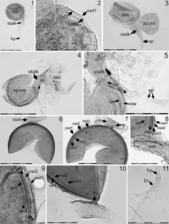
Sporiferous saccule (FIGS. 1–3) is subhyaline to light yellow (to yellow), saccule terminus usually globose (ca. 150–210 mm diam) to subglobose (140–190 3 150–215 mm), with three wall layers (owl1-3) that are in total 2.0–3.5 mm thick (FIG. 2), formed at the end of a hyphal stalk at 30–90 mm from the spore that arises thereafter (FIG. 3). The two outer wall layers (owl1, owl2) are hyaline, evanescent and each 0.4– 1.0 mm thick; the innermost wall layer (owl 3) is subhyaline to light yellow, 1.8–2.7 mm thick (FIG. 2). Only the globose terminus of the sporiferous saccule collapses at the stalk (FIGS. 4, 5) after the spore wall has formed and usually is rapidly detached from the stalk of mature spores in soil samples or pot cultures, while the stalk generally persists on the spore (FIGS. 6, 7). Stalk (5neck) of the sporiferous saccule is yellow- brown to brown, in total 150–450 mm long and arising from a mycelium hypha; 25–40 mm wide at the base of the saccule terminus, tapering to 15–30 mm at the area where the spore arises laterally on the stalk and finally tapering to 5–12 mm toward the hypha (FIGS. 4, 5). Three wall layers of the sporiferous saccule continue over the total length of the stalk with 2.5–4.5 mm at the base of the saccule terminus and at the area where the spore arises, tapering to 1.5–3.0 mm toward the mycelium hypha. The outer layer is hyaline, thin (0.4–1.0 mm) and evanescent; middle layer is subhyaline to light yellow and 1.5–2.5 mm thick at the base of the saccule terminus and at the area where the spore arises, tapering to 0.5–1.0 mm toward the mycelium hypha; this layer is more resistant on the stalk than the outer layer but is often missing on mature spores too; inner layer is yellow- brown to brown, 2.0–3.5 mm thick at the saccule base and at the area where the spore arises (FIGS. 4–8), tapering to 0.5–1.5 mm toward the mycelium hypha (FIGS. 4, 5). One to several septa arising from the brown-pigmented third wall layer separate the con- tents of the sporiferous saccule from the hypha distal to the area where the spore thereafter may arise (FIGS. 3, 5). One to three additional septa may arise from the brown-pigmented wall layer separating the spore contents from the collapsing saccule (FIG. 7) at a later stage of spore formation. Finally an often plug- like, additional septum regularly arises at the spore base that separates the saccule stalk from the developing spore. The thick-walled saccule stalk generally persists on the spore resembling a tangential-lateral, inflated, single-branched subtending hypha (FIGS. 4–8).
Spores found singly in soils or in pot cultures, formed laterally on the hyphal stalk of a sporiferous saccule (FIGS. 4, 5). The spores are yellow-brown to brown, globose (150–200 mm) to subglobose (145– 185 3 175–210 mm) to rarely ovoid or irregular and consist of a yellow-brown to brown outer wall and a hyaline inner wall.
Outer spore wall with four layers (owl1–4; FIGS. 7–9), in total 5–12 mm thick: outer layer owl1 hyaline, thin (0.5–1.0 mm) and evanescent; owl2 subhyaline to light yellow, (2.0–)3.0–4.5(–6.5) mm thick, semipersistent and sometimes slightly expanding in lactic acid based mountants; owl3 yellow-brown to brown (2.5–)4.0–6.0 mm thick, laminated and tightly adherent to owl2; owl4 concolorous with owl3, 0.5–1.5 mm thick and, as tightly adherent to owl3, often difficult to observe. The three outer wall layers are continuous with the wall layers of the stalk of the sporiferous saccule.
Inner wall forms de novo during spore formation and consists of three hyaline layers (iwl1–3; FIGS. 7–10). Outer layer iwl1 is thin and usually hard to detect in PVLG based mountants because it usually is tightly adherent to iwl2; iwl2 is 2.0–3.5 mm thick and finely laminate; iwl3 is 0.5–0.8 mm thick and sometimes difficult to observe, especially when tightly adherent to iwl2; sometimes it readily separates from iwl2 and then is clearly visible forming often several folds (FIG. 10).
Pore at the spore base is (3–)6–11(–14) mm diam; usually occluded by a plug-like septum, which is 1.5–3.5 mm thick and arising directly at the spore base from the laminate owl3 (FIG. 10) and adherent thin layer owl4. The pore at the spore base rarely appears to be open. Then the septa of the saccule stalk— proximal and distal to the terminus of the sporiferous saccule and next to the area where the spore has arisen—might separate the spore contents from the saccule stalk (FIG. 7). In plan view the spore pore resembles a ring or a cicatrix, being connected to the persistent saccule stalk.
Mycelial hyphae (FIG. 11) are hyaline, 1.2–3.0 mm thick with two visible layers when recently formed; outer hyphal layer evanescent; inner hyphal layer persistent and continuous with owl3 of the saccule stalk and the spore.
Formation of arbuscular mycorrhizae so far unknown but assumed from spore ontogeny and morphology, from molecular phylogeny placing the fungus within the Diversisporales and from its molecular detection in the roots.
Etymology. bareai, in honor of Prof. Jose´-Miguel Barea, Spanish pioneer researcher on arbuscular mycorrhizal fungi.
Type. Isolated from the pot culture rhizospheres of Sorghum vulgare and Trifolium pratense s.l. inoculated with soil taken from an endangered plant community of dolomitic endemisms located in the Sierra de Baza (Granada, Spain) at 1600 m a.s.l. (2u979W, 37u379). Holotype and type deposited at Z+ZT (accession number ZT Myc 160); isotypes deposited at OSC (OSC #134502) and GDA-GDAC.
Distribution. So far, O. bareai is known only from the type location in the Sierra de Baza National Park (dolomitic shrub land, Granada, Spain) detected in the rhizosphere of Pterocephalus spathulatus and Thymus granatensis on dolomite.
Molecular analyses.— Sequences of approximately 550 bp corresponding to the NS31-AM1 region of the 18S ribosomal gene were obtained from five single spores. All five sequences were identical. To examine evolutionary relationships among O. bareai and other species in the Glomeromycetes, phylogenetic trees were generated from multiple aligned sequences by using evolutionary parsimony and neighbor joining methods. Because both analyses produced trees with basically the same topology, only the neighbor joining tree is presented (FIG. 12). This topology is largely in agreement with those published by Schüssler et al (2001) and Redecker et al (2007). The phylogenetic analyses suggest that the sequence of O. bareai forms a distinct sister clade to Diversispora spurca and Glomus versiforme sequences of the Glomus group C sensu Schüssler (Schüssler et al 2001, Schwarzott et al 2001) of which so far only the type species Diversispora spurca recently was transferred to a new genus Diversispora of family Diversisporaceae (Walker and Schüssler 2004). The position of O. bareai close to the D. spurca clade was confirmed by PCR amplification and sequencing using the new primer designed for species related to Diversispora spp. (Redecker et al 2007) and DNA extracted from newly formed spores. By using this primer we finally were able to prove the presence of O. bareai in the roots, sampled from the pot cultures.

FIGS. 1–11. Otospora bareai. 1. Terminal sporiferous saccule (sac) with hyphal stalk (5neck) formed on mycelial hypha (hy); bar 5 200 mm. 2. Wall of sporiferous saccule with three wall layers (owl1-3); bar 5 50 mm. 3. Young, immature spore (spore) forming laterally on the stalk of a fully developed terminal saccule (sac). A septum (sp) formed in the hyphal stalk distal to the saccule (sac) and arising spore; bar 5 200 mm. 4. Mature, crushed spore formed on the persistent and pigmented stalk of a collapsing sporiferous saccule (sac); bar 5 200 mm. 5. Mature, crushed spore having outer wall (ow) and inner wall
Species
