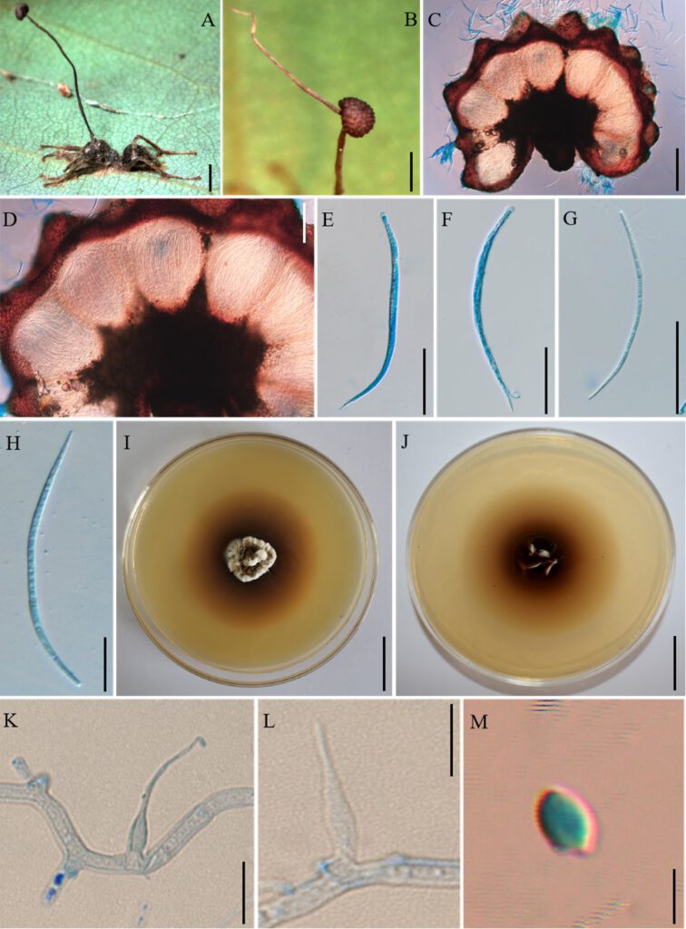
Ophiocordyceps acroascus H. Yu & D.X. Tang.
MycoBank number: MB; Index Fungorum number: IF; Facesoffungi number: FoF 11705;
Description
Subtropical monsoon evergreen broad-leaf forest. Camponotus sp. infected and biting the main vein of a leaf, it was found between 0.5 m and 2 m above the ground. Sexual state: External mycelium produced from the legs and body of the host. Stroma single, and curved at top, produced from dorsal pronotum of the ant, cylindrical, clavate, dark brown at maturity, the top was lighter than other parts of the stroma. Fertile region (ascoma) of lateral cushions produced from the top of the stroma, one to two ascoma were found, hemispherical, brown, averaging 3 × 2–3 mm. Perithecia ovoid, immersed to partially erumpent, with short, exposed neck or rounded ostiole, 247–296 × 190–238 μm. Asci cylindrical, hyaline, curved, thick, 8-spored, 96–197 × 5–8 μm. Ascus caps were hemispherical, prominent and small, 3–5 µm high and 4–6 µm wide. Ascospore vermiform, thin-walled, hyaline, septate, slightly curved to sinuous, round to slightly tapered at apex, 76–113× 2–3 µm. Asexual state:Colonies on PDA slow-growing, 26–27 mm diameter in 60 days at 25 C, milky white and light brown, hard, with protuberant mycelial at the surface, the pigment was produced around colonies, dark brown, reverse light brown to dark brown. Hyphae branched, septate, smooth-walled, hyaline. Hirsutella type-A and Hirsutella type-B were observed. Conidiogenous cell monophialidic, produced from hyphae, smooth, swollen base, cylindrical to lageniform, tapering gradually or abruptly a long neck, slight bending, 6–21 × 1–5 µm. Conidia limoniform, solitary, hyaline, smooth-walled, 2–3 × 1–2 µm.
Material examined: CHINA, biting on leaves of tree seedlings, 18 August 2020, H. Yu (YHH 20121, neotype).
Distribution: Puer City, Yunnan Province, China
Sequence data: LSU: ON555922 (LROR/LR5); SSU: ON555841 (NS1/NS4); EF1a: ON567761 (983F/2218R); RPB1: ON568681(RPB1/RPB1Cr_oph); RPB2: ON568134 (fRPB2-5F/fRPB2-7cR)
Notes: Phylogenetic analyses showed that the O. acroascus formed a sister lineage with O. septa, it was clustered in the O. unilateralis core clade of Hirsutella and was well-supported by bootstrap proportions (BP = 100%) and posterior probabilities (PP = 100%), formed a separate clade with O. septa. O. acroascus was similar to O. septa in the behavior of the host biting a leaf, producing stroma cylindrical, clavate, perithecia immersed and ostiole. However, it differs from O. septa by the ascoma of lateral cushion arising from the top of the stroma, hemispherical, asci curved, ascospore vermiform, slight curved to sinuous, milky white and light brown, producing Hirsutella type-A and Hirsutella type-B phialides, conidiogenous cell monophialidic, cylindrical to lageniform, conidia limoniform. In addition, the size of perithecia, ascoma, asci, ascospore, phialides and conidia also differed from O. septa.

Fig. 2. Ophiocordyceps acroascus (YHH 20121). A: Camponotus sp. infected and biting the main vein of a leaf. B: The ascoma was produced from the stroma. C–D: Cross-section of the ascoma showing the perithecial arrangement. E–F: Asci. G-H: Ascospores. I-J: Colonies on PDA medium. K-L: Conidiogenous cell and conidia. M: Conidia. Scale bars: A = 3000 µm; B = 2000 µm; C = 200 µm; D = 100 µm; E-G = 50 µm; H = 20 µm; I-J = 2 µm; K-L = 10 µm; M = 2 µm.
