Nothotrimmatostroma bifarium (Gadgil & M.A. Dick) Crous, in Crous, Wingfield, Cheewangkoon, Carnegie, Burgess, Summerell, Edwards, Taylor, Groenewald, Stud. Mycol. 94: 207 (2019)
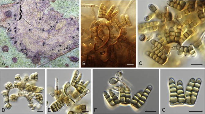
MycoBank number: MB 832034; Index Fungorum Number: IF 832034, Facesoffungi number: FoF 11391 ; Fig. 64.
Basionym: Trimmatostroma bifarium Gadgil & M.A. Dick, New Zealand J. Bot. 21: 49. 1983.
Synonym: Neotrimmatostroma bifarium (Gadgil & M.A. Dick) Quaedvl. & Crous, Persoonia 33: 27. 2014.
Diagnosis: Conidia formed in two basipetal chains, brown, consisting of two parallel, laterally fused rows, with a common thick-ened, transverse base and obtuse apices, 12–24 × 6–14 μm, 6–10-celled when mature (with 3–5 cells per row).
Descriptions and illustrations: Gadgil & Dick (1983), Park et al.(2000).
Description based on CPC 32833: Leaf spots corky, medium brown, irregular, 2– 10 mm diam. Sporodochia hypophyllous, dark brown, in concentric clusters, 150 – 220 μm diam. Co- nidiophores micronematous, branched, septate, medium brown, smooth, densely aggregated, up to 50 μm tall, 3– 4 μm diam. Conidiogenous cells holothallic, integrated, terminal, doliiform to subcylindrical, 9– 11 × 3– 4 μm. Conidia in branched chains, smooth, medium brown, 2– 7-celled, consisting of two parallel rows unequal in length, fused at common thickened base, apices obtuse, (15–)17 – 20(– 22) × (8 –)9(– 10) m, 5– 6-celled in vivo, (13 –)15– 19(– 25) × (7 –)8 – 9(– 10) μm, (2 –)5 – 6(– 7)-celled in vitro.
Culture characteristics: Colonies erumpent, spreading, with sparse aerial mycelium and even, lobate margins, reaching 5 mm diam after 2 wk at 25 °C. On MEA, PDA and OA surface olivaceous grey, reverse iron-grey.
Typus: Australia, New South Wales, Merimbula, on E. dalrympleana, 28 Nov. 2016, P.W. Crous, HPC 1815 (epitype designated here CBS H-24032, MBT388158, culture ex-epitype CBS 143488 = CPC 32833). New Zealand, Kinleith, on E. regnans, Sep. 1981, D.J. Rawcliffe (holotype NZFRI 5, isotype PDD 42845).

Fig. 64. Nothotrimmatostroma bifarium (CPC 32833). A. Leaf spot with conidiomata. B– F. Conidiogenous cells giving rise to conidia. G. Conidia. Scale bars = 10 μm.
