Nothofusarium Crous, Sand.-Den. & L. Lombard, gen. nov. Fig. 8.
MycoBank number: MB 838674; Index Fungorum number: IF 838674; Facesoffungi number: FoF 10986;
Etymology: From the Greek prefix notho-, false, illegitimate; and Fusarium, in reference to the genetic affinity and morphological resemblance to the genus Fusarium s. str.
Type species: Nothofusarium devonianum L. Lombard, Crous & Sand.-Den.
Ascomata unknown. Conidiophores mononematous (aerial conidiophores) or grouped on sporodochia. Aerial co- nidiophores simple, unbranched or irregularly branched, some- times reduced to single lateral phialides or phialidic pegs on the hyphae; conidiogenous cells monophialidic, cylindrical, tapering towards apex, smooth- and thin-walled, with periclinal thickening inconspicuous or absent, solitary. Microconidia not formed. Aerial macroconidia falcate, 1– 5(– 6)-septate, thick-walled, curved to lunate, with a blunt apical cell and often obtuse, poorly- to well- developed foot-shaped basal cell. Sporodochia white, pale luteous to pale citrine. Sporodochial conidiophores irregularly and verticillately branched, consisting of short, smooth- and thin- to thick-walled stipes bearing apical whorls of mono- and poly- phialides. Sporodochial conidiogenous cells monophialidic and polyphialidic, doliiform, subulate to subcylindrical, smooth- and thin-walled, with reduced apical collarette. Sporodochial macroconidia similar to aerial macroconidia. Chlamydospores sub- globose to ellipsoidal, solitary or most commonly in chains.
Diagnostic features: Fusarioid asexual morph characterised by aerial monophialides and sporodochial mono- and polyphialides producing slightly curved and slender, mostly 3-septate macroconidia.
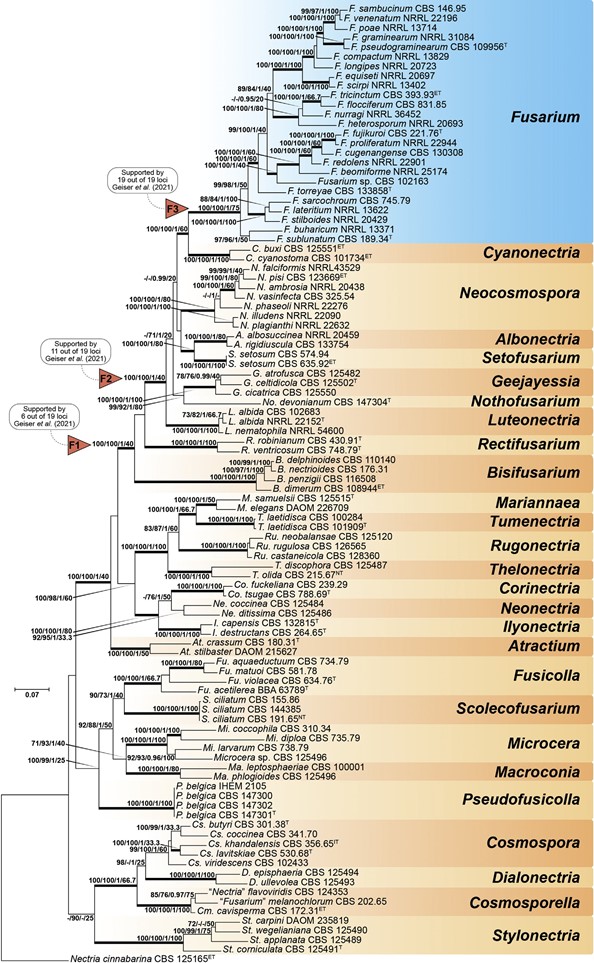
Fig. 7. Maximum-Likelihood (IQ-TREE-ML) consensus tree inferred from the combined ITS, LSU, rpb1, rpb2 and tef1 multiple sequence alignment of members of Nectriaceae. Numbers at the branches indicate support values (RAxML-BS / UFboot2-BS / BI-PP / gCF) above 70 % / 0.95 with thickened branches indicating full support (RAxML-BS / UFboot2-BS / gCF = 100 %; BI-PP = 1). The scale bar indicates expected changes per site. The tree is rooted to Nectria cinnabarina (CBS 125165). Arrows “F1”, “F2” and “F3” indicate the three alternative Fusarium hypotheses sensu Geiser et al. (2013). Ex-epitype, ex-isotype, ex-neotype and ex-type strains are indicated with ET, IT, NT, and T, respectively.
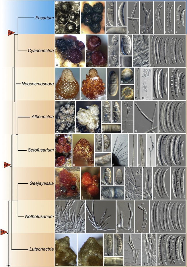
Fig. 8. (Continued).
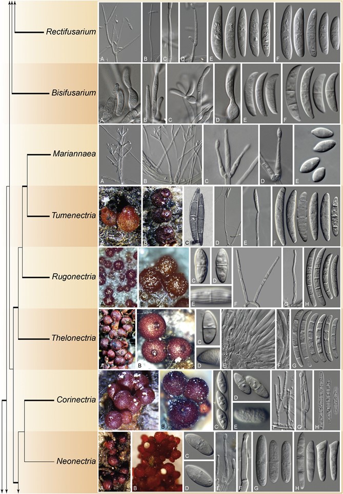
Fig. 8. (Continued).
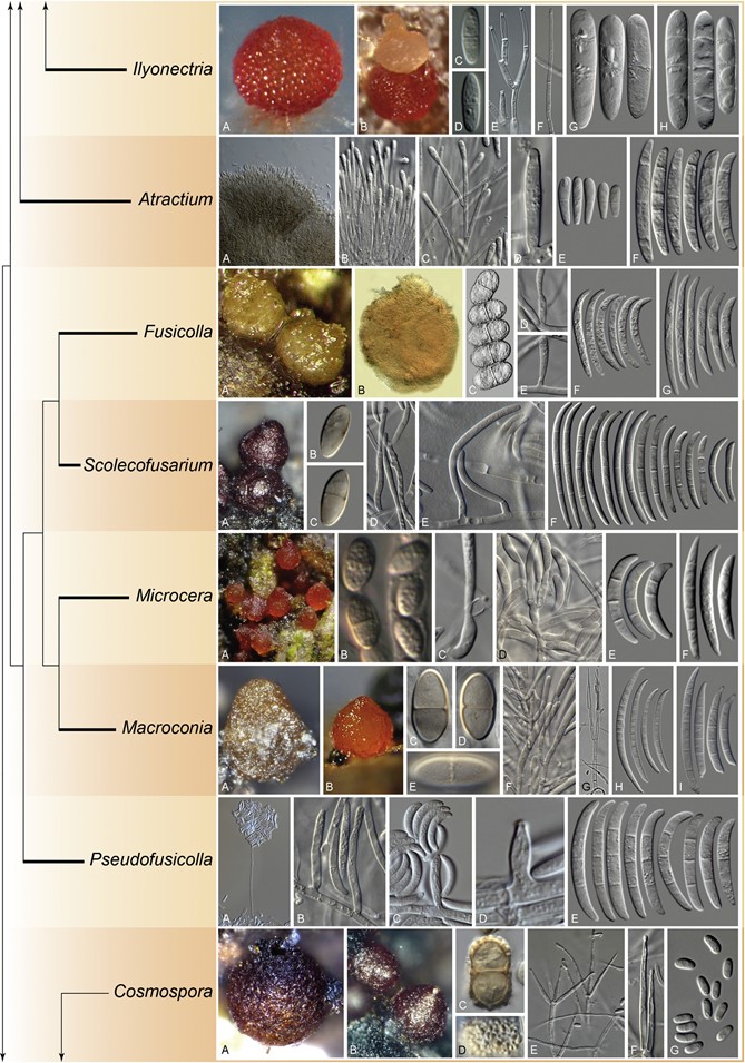
Fig. 8. (Continued).
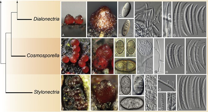
Fig. 8. Morphological features and phylogenetic affinities of fusarioid genera of Nectriaceae and close relatives. The tree was delineated based on the phylogeny presented in Fig. 7 and does not indicate phylogenetic distances. Fully supported branches are indicated in bold. The genus Fusarium is indicated in blue. Arrows “F1”, “F2” and “F3” indicate the three alternative Fusarium hypotheses sensu Geiser et al. (2013). Fusarium. A, B. Ascomata. C– E. Ascospores. F, G. Conidiogenous cells. H– J. Macroconidia. (B. Adapted from Schroers et al. 2011). Cyanonectria. A, B. Ascomata. C–E. Ascospores. F. Conidiogenous cells. G. Macroconidia. Neocosmospora. A, B. Ascomata. C–E. Ascospores. F, G. Conidiogenous cells. H, I. Macroconidia. [A. Adapted from Sandoval-Denis & Crous (2018). G. Adapted from Sandoval-Denis et al. (2019)]. Albonectria. A, B. Ascomata. C–E. Ascospores. F, G. Conidiophores and conidiogenous cells. H. Macroconidia. Setofusarium. A, B. Ascomata. C–E. Ascospores. F– H. Setae formed on sporodochia. I. Conidiophore. J. Conidia. Geejayessia. A, B. Ascomata. C–E. Ascospores. F, G. Conidiophores and conidiogenous cells. H, I. Macroconidia. [A. Adapted from Schroers et al. (2011)]. Nothofusarium. A–D. Conidiophores and conidiogenous cells. E. Conidia. Luteonectria. A, B. Ascomata. C– D. Ascospores. F, G. Conidiophores and conidiogenous cells. H. Conidia. Rectifusarium. A– D. Conidiophores and conidiogenous cells. E, F. Conidia. Bisifusarium. A– D. Conidiophores and conidiogenous cells. E, F. Conidia. Mariannaea. A, B. Conidiophores. C, D. Conidiogenous cells. E. Conidia. Tumenectria. A, B. Ascomata. C. Ascospores. D, E. Conidiophores and conidiogenous cells. F. Conidia. [A– C. Adapted from Salgado-Salazar et al. (2016)]. Rugonectria. A, B. Ascomata. C– E. Ascospores. F, G. Conidiophores and conidiogenous cells. H. Conidia. Thelonectria. A, B. Ascomata. C, D. Ascospores. E, F. Conidiophores and conidiogenous cells. G. Conidia. Corinectria. A, B. Ascomata. C– E. Ascospores. F, G. Conidiophores and conidiogenous cells. H. Conidia. (H. Picture by C. Gonz´alez). Neonectria. A, B. Ascomata. C, D. Ascospores. E, F. Conidiophores and conidiogenous cells. G, H. Conidia. [A. Adapted from Chaverri et al. (2011)]. Ilyonectria. A, B. Ascomata. C, D. Ascospores. E, F. Conidiophores and conidiogenous cells. G, H. Conidia. Atractium. A, B. Conidiophores. C, D. Conidiogenous cells. E, F. Conidia. Fusicolla. A, B. Ascomata. C. Ascospores. D, E. Conidiogenous cells. F, G. Conidia. (A–C. Pictures by C. Lechat). Scolecofusarium. A. Ascomata. B, C. Ascospores. D, E. Conidiophores and conidiogenous cells. F. Conidia. Microcera. A. Ascomata. B. Ascospores. C, D. Conidiogenous cells. E, F. Conidia. (A, B. Pictures by N. Aplin, Fungi of Great Britain and Ireland). Macroconia. A, B. Ascomata. C–E. Ascospores. F, G. Conidiophores and conidiogenous cells. H, I. Conidia. (B. Picture by P. Mlˇcoch). Pseudofusicolla. A, B. Conidiophores and conidiogenous cells. C, D. Conidia. [A– D. Adapted from Triest et al. (2016)]. Cosmospora. A, B. Ascomata. C, D. Ascospores. E, F. Conidiophores and conidiogenous cells. G. Conidia. Dialonectria. A, B. Ascomata. C– E. Ascospores. F, G. Conidiophores and conidiogenous cells. H. Conidia. (A. Picture by P. Mlˇcoch). Cosmosporella. A, B. Ascomata. C–E. Ascospores. F, G. Conidiophores and con- idiogenous cells. H, I. Conidia. (A–E. Pictures by P. Mlˇcoch). Stylonectria. A, B. Ascomata. C– E. Ascospores. F– I. Conidiophores and conidiogenous cells. J. Conidia. (A–C, E. Pictures by B. Wergen).
Species
