Nodulosphaeria senecionis Chethana, Camporesi, E.B.G. Jones & K.D. Hyde.
Index Fungorum number: IF551310; Facesoffungi number: FoF: 00871; Fig. 2
Etymology – The specific epithet senecionis is named after the host Senecio from which the taxon was collected.
Holotype – MFLU 15–1297
Saprobic on dead stems of Senecio spp. Sexual morph: Ascomata 189–275 μm high (including papilla), 257 – 299 μm diam. (x̄ = 238.31 × 284.06 μm, n = 10), scattered, solitary, semi-immersed to superficial, dark brown to black, globose to subglobose, wall rough, with hairs at the base, ostiole central, papillate. Papilla 36 – 47 μm high, 30 – 38 μm diam. (x̄ = 34.1 × 39.9 μm, n = 10), protruding from substrate if immersed, with numerous, brown to dark brown, 25 – 40 μm long setae. Peridium 37 – 59 μm thick at side walls, up to 45 – 79 μm thick near the apex and 36 – 66 μm thick at the base, composed of 4 – 5 layers of thick-walled, dark brown cells of textura globularis. Hamathecium composed of numerous, 2 – 3 μm broad, filiform, anastomosing, septate, pseudoparaphyses, embedded in a gelatinous matrix. Asci 33 – 113 × 4 – 14 μm (x̄ = 82.93 × 10.89 μm, n = 20), 8 – spored, bitunicate, fissitunicate, cylindric – clavate, short pedicellate, apically rounded, with an ocular chamber. Ascospores 30 – 45 × 3 – 5 μm (x̄ = 38.39 × 4.22 μm), overlapping biseriate to tri-seriate, initially hyaline, becoming yellowish brown at maturity, long fusiform to almost cylindrical, enlarged at the third cell from the apex, tapering towards the rounded ends, 8-septate, slightly curved, constricted at the third septum, smooth-walled, with small guttules and small, curved, cylindrical appendages at both ends. Asexual morph: Undetermined.
Material examined – ITALY, Forlì-Cesena Province, Fiumicello-Premilcuore, on dead stem of Senecio sp. (Asteraceae), 12 May 2013, E. Camporesi (MFLU 15–1297, holotype); ibid. (isotype in BBH); on dead stem of Senecio sp., 3 June 2013, E. Camporesi (MFLU 15–1312, paratype); ITALY. Province of Forlì-Cesena, Fiumicello-Premilcuore, on a dead stem of Centaurea sp. (Asteraceae), 8 July 2013, E. Camporesi (MFLU15–1313).
Notes – Based on our phylogenetic analyses (Fig. 1) and morphological comparison, our isolate belongs in Nodulosphaeria in Phaeosphaeriaceae. Nodulosphaeria senecionis is distinct from N. hirta in having larger ascospores. Nodulosphaeria pellita (Fr.) Shoemaker, N. octoseptata (Wehm.) L. Holm, N. derasa (Berk. & Broome) L. Holm and Leptosphaeria senecionis (Fuckel) G. Winter have also been isolated from Senecio sp. and should be compared. Nodulosphaeria pellita and N. octoseptata differ from N. senecionis in having larger ascomata, longer ascospores constricted at the first septum, and with the fourth cell from the apex being enlarged (Shoemaker 1984). Nodulosphaeria derasa differs from N. senecionis in having ascospores, which are constricted at the first septum and with the fourth cell from the apex being enlarged (Shoemaker 1984) . Leptosphaeria senecionis differs from Nodulosphaeria senecionis in having 3-septate, hyaline ascospores (Saccardo 1884). Nodulosphaeria centaureae differs from N. senecionis in having 6 – septate ascospores (Shoemaker 1984).
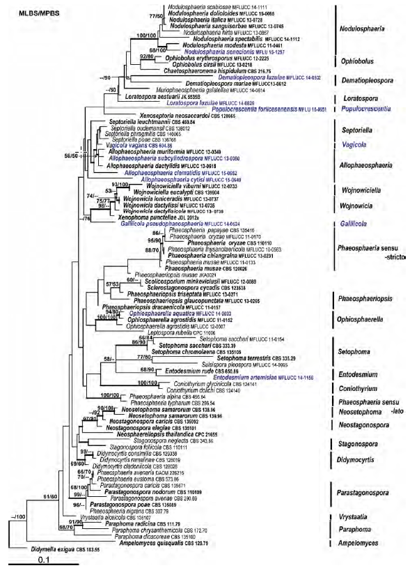
Fig. 1 Phylogram generated from maximum likelihood analysis based on combined LSU and ITS sequenced data of Phaeosphaeriaceae. Maximum parsimony and maximum likelihood bootstrap support values greater than 50 % and Bayesian posterior probabilities greater than 0.75 are near the nodes. The ex-type strains are in bold and the new isolates are in blue. The scale bar indicates 0.1 changes. The tree is rooted with Didymella exigua CBS 183.55.
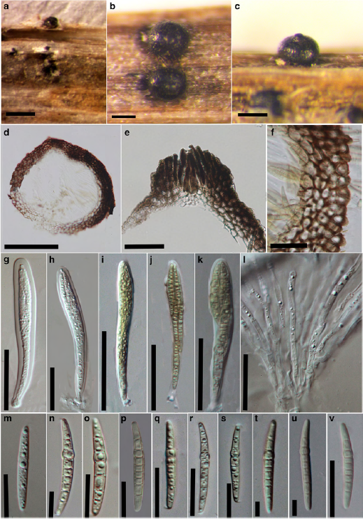
Fig. 2 Nodulosphaeria senecionis (holotype) a, b Appearance of ascomata on the host substrate c Close up of ascoma d Section of the ascoma e Section of the ostiole with setae f Peridium g, h Immature asci i – k Mature asci l Hamathecium of septate, cellular pseudoparaphyses m Immature hyaline ascospore n – v Mature hyaline to yellowish brown ascospores. Scale bars: a = 500 μm, b, c = 200 μm, d = 100 μm, e = 25 μm, f, m – v = 10 μm, g – k = 20 μm, l = 5 μm.