Neosonderhenia eucalypti Crous, sp. nov.
MycoBank number: MB 832026; Index Fungorum number: IF 832026; Facesoffungi number: FoF 15387. Fig. 57.
Etymology: Name refers to Eucalyptus, the host genus from which this fungus was isolated.
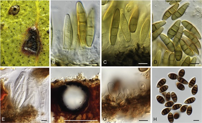
Leaf spots dark brown, amphigenous, subcircular, 1–3 mm diam, with prominent red-purple margin. Conidiomata separate, pycnidial, brown, globose, 200–350 μm diam with central ostiole; wall of 6–8 layers of brown textura angularis. Conidiophores reduced to conidiogenous cells lining the inner cavity, hyaline to pale brown, smooth to verruculose, 5–12 × 5–7 μm, proliferating percurrently at apex. Paraphyses hyaline, hypha-like, septate, 3–4 μm diam, up to 60 μm long, intermixed between conidiophores. Conidia solitary, brown, verruculose, fusoid-ellipsoid, 3-distoseptate with central septal pore in each septum, apex subobtuse, base trun- cate, 3–4 μm diam, (27–)32–36(–40) × (8–)9(–10) μm.
Culture characteristics: Colonies erumpent, spreading, surface folded, with moderate aerial mycelium and smooth, lobate margin, reaching 10 mm diam after 2 wk at 25 °C. On MEA, PDA and OA surface dirty white, reverse ochreous.
Typus: Australia, New South Wales, Mildura, Mungo National Park, on E. costata, 28 Aug. 2015, B.A. Summerell, HPC 2229 (holotype CBS H-24041, culture ex-type CBS 145081 = CPC 34405); idem., CBS 145082 = CPC 34395.
Notes: Other than Neosonderhenia foliorum, N. eucalypti should also be compared to Sonderhenia, which presently includes two species, S. eucalyptorum (conidia 40– 48 × 5– 6 μm; Hansford 1954) and S. eucalypticola (conidia 20– 26 × 9– 11 μm; Swart & Walker 1988), both associated with small, circular leaf spots on Eucalyptus. Swart & Walker (1988) treated Hendersonia fraserae (conidia 23– 28 × 6– 9 μm; Hansford 1954) as synonym of Hendersonia eucalypticola, which was placed in a new genus as Sonderhenia eucalypticola.

Fig. 57. Neosonderhenia spp. A–D. N. eucalypti (CBS 145081). A. Leaf spot. B– D. Conidiogenous cells and conidia. E– H. N. foliorum [K(M) 251545]. E. Ascus with ascospores. F. Transverse section through conidioma. G. Conidiogenous cells. H. Conidia. Scale bars: F = 250 μm, all others = 10 μm.
