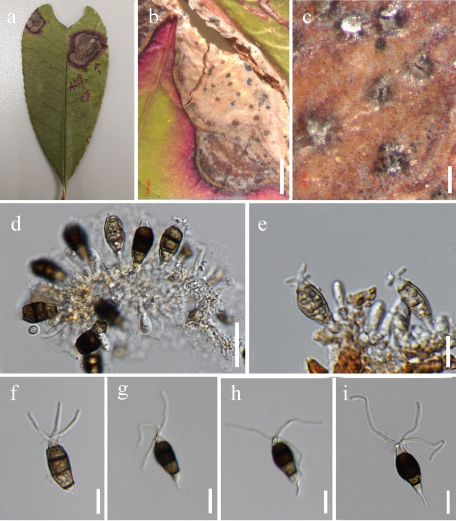
Neopestalotiopsis photiniae Y.R. Sun & Yong Wang bis, in Sun, Jayawardena, Sun & Wang, Microbiology Spectrum: e03987-22, 6 (2023)
Index Fungorum number: IF 571231; MycoBank number: MB 571231; Facesoffungi number: FoF 12914;
Etymology – Referring to the host plant from which the fungus was isolated.
Holotype – HKAS 125895
Associated with leaf spots of Photinia serrulat. Symptoms irregular shape, pale to brown, slightly sunken spots appear on the leaves of Photinia serrulata, which later expand outwards. Small spots gradually enlarged, changing to brown circular ring spots with a dark brown border. Sexual morph: Not observed. Asexual morph: Conidiomata solitary, subglobose to globose, unilocular, dark brown, semi-immersed on leaves. Conidiophores indistinct, often reduced to conidiogenous cells. Conidiogenous cells sub-cylindrical, ampulliform, hyaline. Conidia 20 to 29 × 5 to 12 μm (x̄ = 23 × 9 μm, n = 40), L/W ratio of 2.6, broadly fusiform, straight to slightly curved, 4-septate; basal cell obconic with a truncate base, hyaline to pale brown, 1 to 5 μm long; three median cells 13 to 19 μm long (x̄ = 16 μm, n = 40), brown to dark, wall rugose, versicolorous; second cell from base pale brown to brown, 4 to 6 μm long; the third and fourth cells, dark brown to black, are not easily distinguished, septate indistinct, 10 to 13 μm long; apical cell 2 to 4 μm long, hyaline, conic to acute; with 2 to 3 tubular appendages on the apical cell, inserted at different loci in a crest at the apex of the apical cell, unbranched, 17 to 33 μm long; single basal appendage, unbranched, tubular, centric, 1 to 6 μm long.
Culture characteristics – Conidia germinated on PDA within 12 hours at 25 ℃ from single-spore isolation. Apical cells produced germ tubes. Colony diameter reached 80 mm after three weeks at 25 ℃ on PDA media, circular, surface rough, flat, white from above and below.
Material examined – China, Guizhou Province, Guiyang City, Nanming District, Xiaochehe Road, Guiyang Ahahu National Wetland Park, on leaf spots of Photinia serratifolia (Rosaceae), 21 September 2019, Y.R. Sun, AH9 (HKAS 125895, holotype); ex-type culture, MFLUCC 22-0129; ibid., on leaf spots of Photinia sp. (Rosaceae), 21 September 2019, Y.R. Sun, AH9-1, living culture, GUCC 21-0820.
Notes – Neopestalotiopsis photiniae is phylogenetically related to N. sichuanensis and N. vheenae (Fig. 1). Neopestalotiopsis photiniae differs by its thinner conidia (L/W ratio = 2.7 vs. L/W ratio = 4.1) from N. sichuanensis (Jiang et al. 2021). Neopestalotiopsis photiniae is morphologically indistinguishable from N. vheenae (Prasannath et al. 2021). However, they have 13 base pairs (without gap, 473 bp) in the TEF region. The result of the PHI test showed there is no obvious recombination (Фw = 1.0) between N. photiniae and its closely related taxa (Fig 2a). Therefore, N. photiniae is introduced as a new species.

Figure 6 – Neopestalotiopsis photiniae (HKAS 125895, holotype). a Host. b Leaf spot on Photinia serrulata. c Close up view of conidiomata. d, e conidia attached to conidiogenous cells. f–i Conidia. Scale bars: d = 1000 µm, c = 200 µm, d = 20 µm, f–i = 10 µm.
