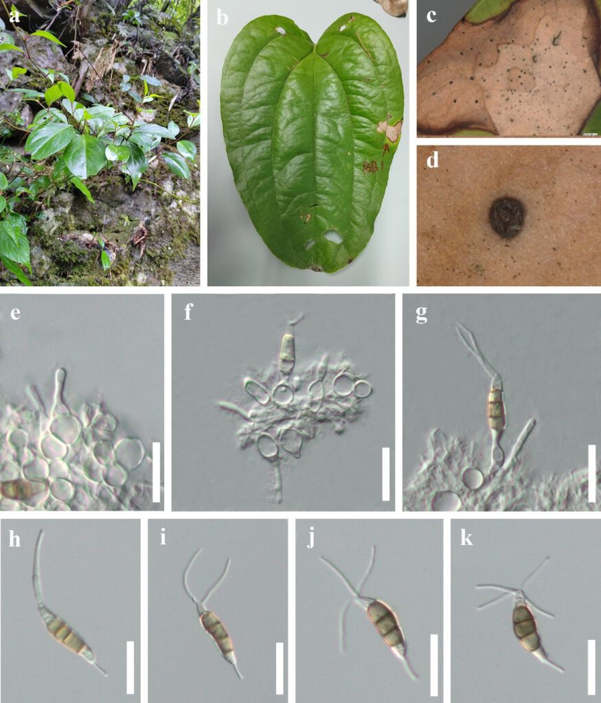
Neopestalotiopsis chinensis Y.R. Sun &Yong Wang bis, sp. nov. Fig. 4
Index Fungorum number: IF; MycoBank number: MB; Facesoffungi number: FoF 06683;
Etymology – The specific epithet is referring to China, the country from where the taxa were isolated.
Holotype – HKAS 124560
Associated with leaf spot of Cyrtomium fortune, Lithocarpus sp. and Smilax scobinicaulis. Symptoms irregular shape, pale brown, small spots gradually enlarged, changing to brown circular ring spots with a dark brown border. Sexual morph: Not observed. Asexual morph: Conidiomata solitary, subglobose to globose, unilocular, dark brown, semi-immersed on leaves. Conidiophores indistinct, often reduced to conidiogenous cells. Conidiogenous cells subcylindrical or ampulliform, hyaline. Conidia 21.0–31.0 × 4.0–7.0μm, ± SD = 26.5 ± 2.3 × 6.0 ± 0.7 μm (n = 30), L/W ratio = 4.4, fusiform, straight to slightly curved, 4-septate; basal cell obconic with a truncate base, hyaline, 3.0–6.8 μm long ( ± SD = 5.2 ± 0.85 μm); three median cells doliiform to cylindrical, 11.5–18.0 μm long ( ± SD = 15.5 ± 1.4 μm), yellow to brown, concolourous, septa darker than the rest of the cell; second cell from base yellow to brown, 3.5–6.5 μm long ( ± SD = 5.0 ± 0.6 μm); third cell yellow to brown, 3.5–6.5 μm long ( ± SD = 5.0 ± 0.64 μm); fourth cell yellow to brown, 4.0–6.5 μm long ( ± SD = 5.0 ± 0.9 μm); apical cell 2.5–6.0 μm long ( ± SD = 4.0 ± 0.7 μm), hyaline, conic to acute; with 1–4 tubular appendages on apical cell, inserted at different loci in a crest at the apex of the apical cell, unbranched, 13.0–26.0 μm long ( ± SD =19.5 ± 3.2 μm); single basal appendage, unbranched, tubular, centric, 2.5–7.0 μm long ( ± SD = 4.5 ± 1.1 μm).
Culture characteristics – Conidia germinated on PDA within 12 hours at 25 ℃ from single-spore isolation. Apical cells produced germ tubes. Colony diameter reached 80 mm after two weeks at 25 ℃ on PDA media, circular, surface rough, flat, white from above, yellow from below.
Material examined – China, Guizhou Province, Qiannan Buyi and Miao Autonomous Prefecture, Libo District, leaf spot of Smilax china L.(Liliaceae), 12 March 2022, Y.R. Sun, bb1 (HKAS 124560, holotype); ex-type-living cultures, GUCC 807; China, Guizhou Province, Tongren City, Jiangkou District, Yamugou Parkland, leaf spot of Lithocarpus sp.(Fagaceae), 20 May 2022, Y.R. Sun, JK15-2 (HKAS 124565, Paratype); living cultures, GUCC 808; China, Guizhou Province, Guiyang City, Baiyun District, Changpoling National Forest Park, leaf spot of Dryopteris crassirhizoma Nakai (Dryopteridaceae), 20 August 2021, Y.R. Sun, CL1-2 (HGUP 22-xxx, dried culture); living cultures, GUCC 813.
Notes – Three strains of Neopestalotiopsis chinensis (GUCC 807, GUCC 813 and GUCC 808) have identical ITS, TEF, and TUB sequences to isolates CFCC-54337 and ZX12-1, which were previously provided by Jiang et al. (2021). However, they did not introduce it as a new species due to the lack of neighboring species to compare the morphology. In this study, GUCC 807, GUCC 808 and GUCC 813 have the same morphology. However, they have longer conidia than CFCC-54337 and ZX12-1 (21.0–31.0 × 4.0–7.0 vs. 19.9–23 ×5.8–7.6). In phylogenetic analyses, N. chinensis is close to N. formicarum, N. photiniae N. sichuanensi and N. vheenae. The PHI test on N. chinensis indicated that there is no significant recombination (Фw = 1.0) between N. chinensis and its closely related taxa. Thus, we introduce N. chinensis as a new species and assign GUCC 807 as the holotype, due to CFCC-54337 and ZX12-1 are invalid. Neopestalotiopsis chinensis appears to be a common phytopathogen as it has been found in leaf spots on different plants.

Figure 4 – Neopestalotiopsis chinensis (HUGP 22-xx, holotype). a Host. b Leaf spot on Smilax scobinicaulis. c, d Close up view of conidiomata. e–g Conidia attached to conidiogenous cells. h–k Conidia. Scale bars: e–k = 20 µm.
