Neoleptosporella camporesiana R.H. Perera & K.D. Hyde, new host record (Dhanu) (Tak33)
MycoBank number: MB 556898; Index Fungorum number: IF 556898; Facesoffungi number: FoF 06962; Fig. **
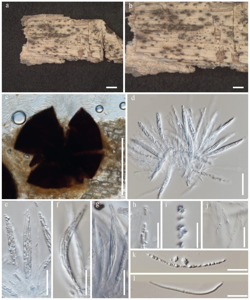
Saprobic on a dead leaves of Dracaena sp. Sexual morph: Appearing as black spot raised dome-shaped, with a central short papilla. Ascomata 250–350 μm high, 500–600 μm diam. (x̅= 300 × 550 μm, n = 20), solitary or aggregated, immersed beneath small clypeus, subglobose to depressed globose. Paraphyses 10–15 μm diam. (x̅= 13 μm, n = 10), hyaline, branched, septate. Asci 150–200 × 10–15 μm (x̅= 180 × 13 μm, n = 20), 8-spored, unitunicate, cylindrical, long pedicellate, apex rounded with a wedge-shaped, J-, apical ring. Ascospores 100–150 × 2.5–4 μm (x̅= 130 × 3 μm, n = 20), fasciculate, parallel becoming spiral at maturity, filiform, straight or curved, hyaline, aseptate, rounded at the apex, pointed at the base, smooth-walled, without appendages. Asexual morph: Undetermined.
Material examined – THAILAND, Tak Province, on dead leaves of Dracaena sp., 21 August 2019, Napalai Chaiwan, Tak33 (MFLU ****, holotype).
Host – Dracaena sp.—(This study).
Distribution – Thailand—(This study).
GenBank accession numbers – ITS = ****, LSU = ****.
Notes – In the phylogenetic analysis of Chaetosphaeriales taxa, our novel strain (MFLU***) nested with Neoleptosporella species and this clade is supported with 71% in ML. Within this clade the new isolates constituted a strongly supported (100% ML, Fig ***) monophyletic lineage with Neoleptosporella camporesiana (MFLUCC 15–1016). Our strain was found on Dracaena sp. (Tak, Thailand) whereas the holotype of N. camporesiana was found on a dead branch of an unidentified plant in Chiang Rai, Thailand). Based on the morphological similarity and phylogenetic evidences, we account our new collection as another report of Neoleptosporella camporesiana. The other remaining member of this genus is Neoleptosporella clematidis and this was reported from Clematis subumbellata in Chiang Rai, Thailand (Phukhamsakda et al. 2020).

Figure ***– Neoleptosporella camporesiana (MFLU ***, herbarium). a Herbarium material. b Appearance of ascomata on host substrate. c A squashed ascoma. d–h Asci (h in Melzer’s reagent). i, k–l Ascospores. j Paraphyses. Scale bars: a–b = 1000 μm, c = 50 μm, d, e, f, g = 20 μm, h–l = 10 μm.
