Neokamalomyces indicus Sanjay & Raghv. Singh, in Yadav, Verma, Singh, Singh, Chaurasia, Singh & Kumar, Phytotaxa 571(2): 156 (2022)
Index Fungorum Number: IF 843768; Facesoffungi Number: FoF 15718
Etymology – ‘indicus’, referring to India, the country where the fungus was discovered.
Type – India, Uttarakhand, Haridwar, Har Ki Pauri, 29.9567°N 78.1710°E, on living leaves of Ficus benghalensis L. (Moraceae), July 2019, coll. Sanjay Yadav, holotype (AMH 10233), isotype (MH-BHU 13), ex-type living culture (NFCCI 4870).
Diagnosis – Differs from Parapallidocercospora colombiensis by its presence of only internal mycelium, epigenous colonies, and conidiomata pycnidial type, conidiophores develop from the inner lining of the conidiomatal wall, hyaline, shorter and reduced to conidiogenous cells, conidia hyaline to light olivaceous and always smooth.
Leaf spots numerous, amphigenous, circular to subcircular, brown, with a dark brown border with a greyish white centre (with many blackish dots representing conidioma), 5–10 mm diam, later on, became irregular and covered the entire leaf surface (Fig. 1a–e). Asexual morph: Conidiomata pycnidial, brown to darkish brown, subepidermal, epigenous, visible on upper sides of the leaf, several in each lesion, immersed to erumpent, subglobose to globose, 49–180 µm diam., with a central ostiolum, releasing a hyaline conidial mass; conidiomatal wall without distinctly differentiated layers of textura angularis, the outer cells with brown, somewhat thickened walls, the inner cells hyaline, thin-walled (Fig. 3a–d). Ostiole single, circular, central (Fig. 3a–d). Conidiophores hyaline, reduced to conidiogenous cells, line the inner cavity (Fig. 3a–e). Conidiogenous cells hyaline, tightly aggregated, cylindrical and tapering gradually toward the apex, ampulliform or lageniform with a relatively long neck, holoblastic, proliferating sympodially, smooth, percurrent proliferations not observed, 8–12(–16) × 3–7 μm, scars unthickened (Fig. 3e–g). Conidia cylindrical, weakly to strongly curved, or flexuous, gradually attenuated to a rounded apex, gradually or more abruptly attenuated into a broadly truncate base, 0–4-septate, not or indistinctly constricted around the septa, hyaline to light olivaceous, (10–)23–36(–50) × (1.5–)2-4(–5) μm, hila unthickened to slightly thickened (Fig. 4a–m). Sexual morph: not seen.
Description in vitro – Only sterile mycelium was found without developing any kind of fruiting body or spores (Fig. 2k).
Culture characteristics – Colonies on PDA restricted and erumpent, surface folded, cerebriform to irregularly pustulate, mostly covered by a dense woolly floccose mat of smoke grey aerial mycelium, dark blackish green, reverse fuscous-black, with an irregular margin, reaches 8 mm diam in 28 days at 25 °C with an even to slightly ruffled and glabrous margin (Fig. 2a–j).
Note – Present collections share a superficial morphological resemblance with Septoria and septoria-like fungi (Crous et al. 2009a, b) due to the presence of pycnidial-type conidiomata that are internally lined by conidiophores. However, based on multi-gene analyses, it is quite distant from the Septoria and septoria-like clade. Phylogenetic analyses based on the LSU, RPB2 and ITS sequence data retrieved from the ex-epitype culture of different genera of Mycosphaerellaceae along with Septoria and septoria-like fungi were chosen to establish the exact phylogenetical position of the present collection. Neokamalomyces indicus is morphologically indistinguishable from previous broad concepts of Septoria s. lat., just based on morphology. However, the phylogenetic position of N. indicus is quite distant from the Septoria s. lat. clade, which now allows the retention of this species in the latter genus. These results justify the introduction of a new genus for this lineage, viz., Neokamalomyces.
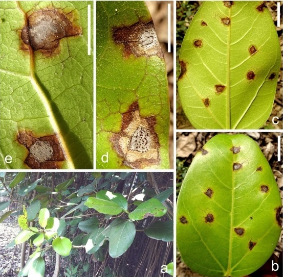
Figure 1 – Neokamalomyces indicus on Ficus benghalensis. a Host plant in natural habitat, b Initial stage of symptom on the upper surface of the leaf, c Initial stage of symptom on the lower surface of the leaf, d, e Conidiomata on host tissue. Bars: b, c = 20 mm, d, e = 10 mm.
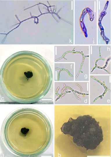
Figure 2 – Neokamalomyces indicus on PDA (NFCCI 4870, ex-type culture). a, b Colony on PDA front view, c Colony on PDA reverse view, d–h Germinated conidia in water droplet in cavity slide after 12-15 hours, i, j Germinated conidia on PDA (stained with cotton-blue), k Development of mycelia on PDA. Bars: a = 10 mm, b = 5 mm, c = 10 mm, d–h = 20 μm, i–k = 10 μm.
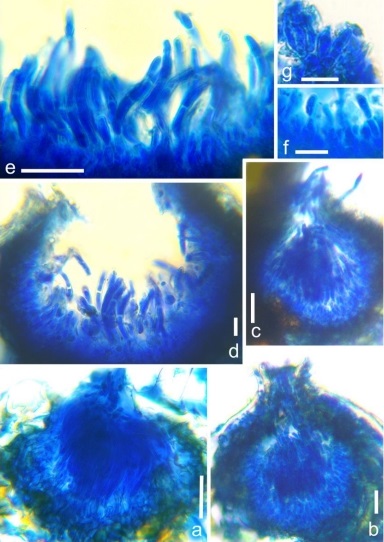
Figure 3 – Fruiting body of Neokamalomyces indicus (holotype, AMH 10233). a–d Vertical section through conidioma, e, f Conidia with conidiophores, g Conidiogenous cells. Bars: a–e = 20 μm, f, g = 10 μm.
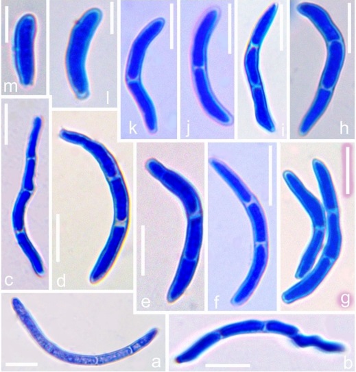
Figure 4 – Conidia of Neokamalomyces indicus (holotype, AMH 10233). Bars: a–k = 10 μm, l, m = 5 μm.
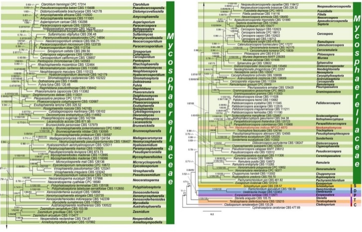
Figure 5 – Consensus phylogram (50% majority rule) resulting from a maximum likelihood of the combined three-genes (LSU, RPB2 and ITS) sequence alignment. The Bayesian posterior probabilities (≥ 0.50; BI-PP), maximum likelihood bootstrap support values (≥ 50%; ML-BS) and maximum parsimony bootstrap support values (≥ 50%; MP-BS) are given at the nodes (BI-PP/ML-BS/MP-BS). Red names indicate Neokamalomyces indicus. A vertical bar is used to the right of the coloured boxes, encompassing all genera within their respective families. The family name Mycosphaerellaceae is unabbreviated, while the rest are abbreviated as follows: D = Dissoconiaceae, P = Phaeothecoidiellaceae, S = Schizothyriaceae, T = Teratosphaeriaceae, C = Cladosporiaceae. The tree was rooted to Cylindroseptoria ceratoniae (CBS 477.69).