Neokalmusia didymospora D.Q. Dai & K.D. Hyde, in Dai, Bahkali, Ariyawansa, Li, Bhat & Chukeatiro, Sydowia 68: 20 (2016)
Index Fungorum number: IF 550836; MycoBamk number: MB 550836; Facesoffungi number: FoF 00061
Saprobic on dead culms of Microstegium sp. Sexual morph: Ascomata 420–460 µm high, 140–220 µm diameter, immersed and raising host tissue, becoming erumpent, solitary, scattered, globose, brown to dark brown, ostiolate, wide, brown to reddish brown, smooth. Peridium 20–35 µm, wider at the apex and thinner at the base, composed of 4–6-layers of dark brown, cells of textura angularis, cells towards the inside lighter, at the outside, darker, fused with the host tissues. Hamathecium comprising numerous, 1.5–1.8 µm wide, filamentous, branched, septate, pseudoparaphyses. Asci 135–140 × 8–9.5 µm, 8-spored, bitunicate, fissitunicate, elongate-clavate to short cylindrical, long pedicellate, thick-walled at the apex, with a minute ocular chamber. Ascospores 12–13.5 × 5–5.5 µm, overlapping 1–2-seriate, hyaline to light brown when immature, becoming brown to blackish brown when mature, ellipsoidal, unequally 1-septate and strongly constricted at the septum, with the upper cell wider and pointed and lower cell longer and rounded, smooth-walled, thick-walled, without a mucilaginous sheath.
Known distribution (based on molecular data) – Thailand, Chiang Rai (Dai et al. 2015), China, Yunnan, Kunming (Hyde et al. 2020).
Known hosts (based on molecular data) – Bamboo (Dai et al. 2015), Microstegium sp. (Poaceae) (Hyde et al. 2020)
Material examined – China, Kunming, KIB premises, on decaying stems of Microstegium sp. (Poaceae), 20 October 2016, A. Karunarathna, AKKIB 48 (MFLU 17–0371, KUN 97364, new host and geographic record); living culture, MFLUCC 17–381; KUMCC 16–0233.
GenBank number – LSU: MN989184.
Notes – Our strain is phylogenetically related to N. didymospora (Fig. 2) and both are morphologically similar. Neokalmusia didymospora is characterized by immersed, orange to brown, thin-walled ascomata, bitunicate, fissitunicate, cylindrical asci with long pedicel and brown, 1-septate ascospores. This fungus shares similar characters with N. brevispora and N. scabrispora in having immersed ascomata, a thin-walled peridium containing host and fungal tissue and verrucose ascospores (Tanaka and Harada, 2004; Ariyawansa et al., 2014). However, N. didymospora differs from N. brevispora and N. scabrispora in ascomata forming an orange to brown, flat, clypeus on the host surface,long-pedicellate asci and 1-septate ascospores.
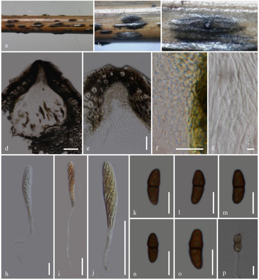
Fig. 1– Neokalmusia didymospora (MFLU 17–0371, new host and geographic record). a–c Appearance of ascoma on host. d Longitudinal section of ascoma. e Longitudinal section of ostiole. f Peridium. g Pseudoparaphyses. h–j Asci. k–o Ascospores. p Germinating ascospore. Scale bars: d = 50 µm, e = 50 µm, f = 10 µm, g = 5 µm, h–j = 20 µm, k–p =10 µm.