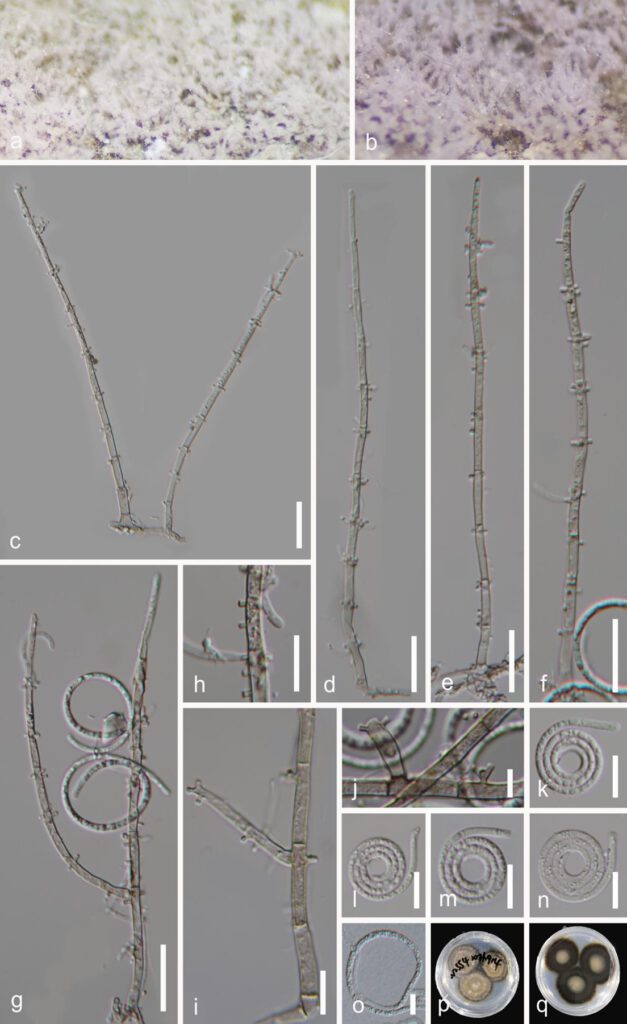
Neohelicomyces hainanensis Y.Z. Lu & J.C. Kang, sp. nov. Fig. 4
MycoBank number: MB; Index Fungorum number: IF; Facesoffungi number: FoF 13103;
Holotype: GZAAS 22-2009
Etymology: ‘hainanensis’ referring to collecting site.
Saprobic on decaying wood. Sexual morph: Undetermined. Asexual morph Hyphomycetous, helicosporous. Colonies on the substratum superficial, effuse, gregarious, white to pink. Mycelium partly immersed, hyaline to pale brown, septate, with masses of crowded, glistening conidia. Conidiophores 137–197 μm long, 2.5–5 μm wide ( = 170 × 4 µm, n = 30), macronematous, mononematous, erect, septate, sparsely branched, pale brown, arising directly on substrate, hyaline to pale brown, smooth-walled. Conidiogenous cells 11–17 × 3–4 µm ( = 14 × 3.5 µm, n = 30), holoblastic, mono- to polyblastic, integrated, cylindrical, with lateral minute denticles (1–2 μm long, 1–1.5 μm wide). Conidia 14–21 µm in diam., 1.5–3 µm wide ( = 17 × 2 µm, n = 30), conidial filament 82–136 µm long, solitary, acropleurogenous, helicoid, coiled 21/2–33/4 times, becoming loosely coiled in water, rounded at tip, guttulate, indistinctly multi-septate, hyaline to yellowish, smooth-walled.

Figure 4. Neohelicomyces hainanensis (GZAAS 22-2009, holotype). a, b Colony on decaying wood. c–g Conidiophores and conidia. h–j Conidiogenous cells. k–n Conidia. o Germinating conidium. p, q Colonies on PDA from above and below. Scale bars: c–g = 20 µm, h–i, k–n = 10 µm, j = 5 µm
