Neodiluviicola W. Dong & H. Zhang, gen. nov.
Index Fungorum number: IF558080; Facesoffungi number: FoF 09565
Etymology: – in reference to the morphologically similar genus Diluviicola
Saprobic on submerged bamboo or wood.
Sexual morph: Ascomata scattered or solitary, semi-immersed to superficial, subglobose or ellipsoidal, black, with a lateral, short neck. Necks black, lying horizontally on the host substrate or curving upwards. Peridium thin, comprising several layers of dark brown to black, compressed cells of textura angularis. Paraphyses numerous, cylindrical, unbranched, hyaline, septate, tapering towards the apex. Asci 8-spored, unitunicate, clavate to subcylindrical, pedicellate, with a distinct, refractive, wedge-shaped, apical ring. Ascospores obliquely uni-seriate or partially biseriate or sometimes triserial, fusiform, straight or slightly curved, aseptate or occasionally 1-septate, hyaline, thin-walled, with bipolar filamentous appendages. Appendages initially coiled in the bipolar chambers of ascospores then unfurl from within the chambers after releasing.
Asexual morph: Undetermined.
Type species: – Neodiluviicola aquatica (W. Dong et al.) W. Dong & H. Zhang
Notes: – Diluviicola aquatica was introduced based on its morphological similarities with the type species D. capensis in having black ascomata with a short, lateral neck and the same unfurling mechanism of ascospore appendages (Hyde et al. 1998, Zhang et al. 2017). However, D. aquatica is phylogenetically distant from D. capensis and forms a basal branch to Cataractispora and Pseudoproboscispora (Fig. 1). We notice that D. aquatica and D. capensis have different unfurling mechanisms of appendages. The appendages of D. aquatica initially coiled in the chambers of ascospores then unfurl from within the chambers after their release (Zhang et al. 2017). In contrast, the appendages of D. capensis are coiled in the conical cap which detached from the tip of ascospores once released in water and unfurl from within the cap (Hyde et al. 1998, this study). In addition, D. aquatica has clavate asci, while D. capensis has cylindrical asci.
Pseudoproboscispora has a similar appendage unfurling mechanism that is initially coiled and then unfurls in water to form long threads (Wong et al. 1999a), but lacks bipolar chambers containing coiled appendages. Instead, its appendages are initially proboscis-like, which also differ from D. aquatica. Therefore, we establish a new genus Neodiluviicola to accommodate D. aquatica. Different unfurling mechanisms of ascospore appendages of three genera and one species in Pseudoproboscisporaceae are summarized in Table 3.
Table 3 Different unfurling mechanisms of ascospore appendages of Cataractispora receptaculorum, Diluviicola, Neodiluviicola and
Pseudoproboscispora
| Taxa | Unfurling mechanisms | References |
| Cataractispora receptaculorum
|
Initially in the form of mucilaginous pads and then unfurl readily in water to form long threads | Ho et al. 2004
|
| Diluviicola | Initially coiled in the conical cap which detached from the tip of ascospores once released in water and unfurl from within the cap | Hyde et al. 1998, this study
|
| Neodiluviicola | Initially coiled in the chambers of ascospores then unfurl from within the chambers after releasing | Zhang et al. 2017 |
| Pseudoproboscispora | Initially coiled as proboscis-like and then unfurl in water to form long threads | Wong et al. 1999a, Zhang et al. 2017 |
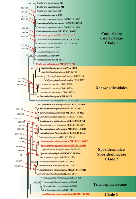
Figure 1 – RAxML tree generated from combined LSU, ITS, TEF and RPB2 sequence data. Bootstrap support values for maximum likelihood (the first value) equal to or greater than 75% and Bayesian posterior probabilities (the second value) equal to or greater than 0.95 are placed near the branches as ML/BYPP. The tree is rooted to Aureobasidium pullulans CBS 584.75 and Dothidea insculpta CBS 189.58. The ex-type strains are indicated in bold and newly generated sequences are indicated in red. The new species introduced in this study are underlined.
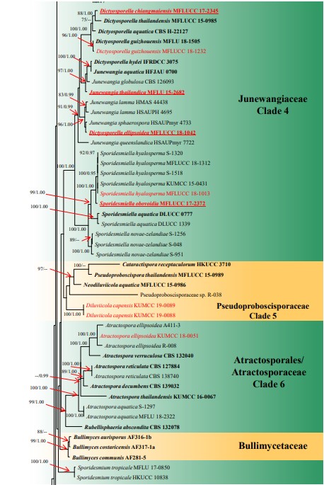
Figure 1 – Continued.
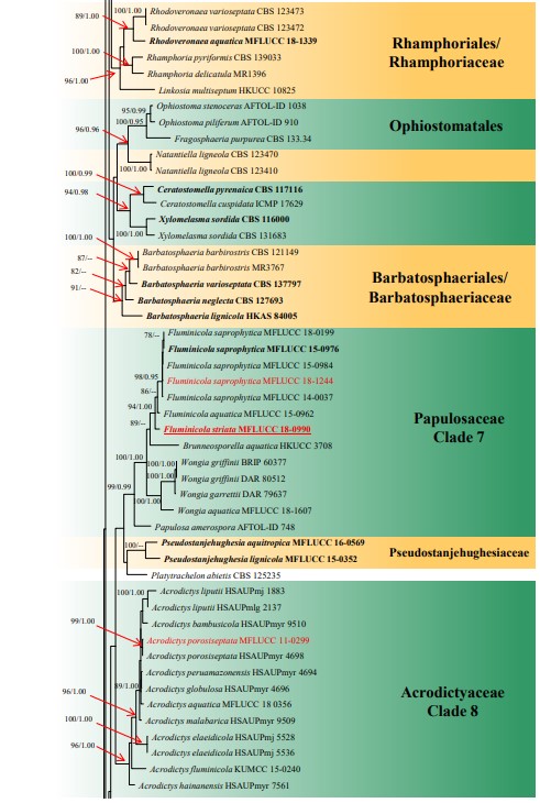
Figure 1 – Continued.
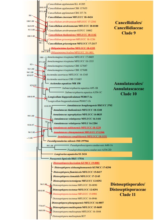
Figure 1 – Continued.
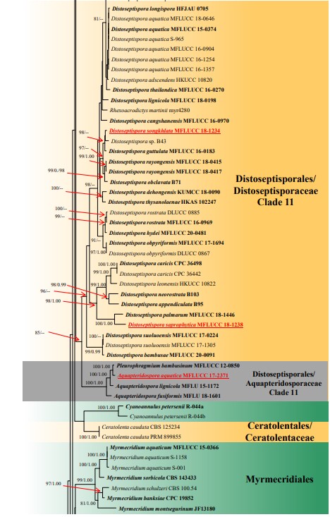
Figure 1 – Continued.
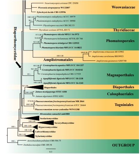
Figure 1 – Continued.
Species
