Neocercosporella peristrophes (Syd.) Sanjay Yadav & Raghv. Singh, in Yadav, Singh, Verma, Singh & Kushwaha, Mycol. Progr. 22(4, no. 26): 10 (2023)
Index Fungorum Number: IF 840501, MycoBank Number: MB 840501, Facesoffungi number: FoF 15861
Basionym: Cercosporella peristrophes Syd., Ann. Mycol. 31: 93 (1933).
Synonyms: Cercosporella peristrophes var. microspora N.D. Sharma & R.P. Mishra, J. Indian Bot. Soc. 56: 133 (1977).
Pseudocercosporella andrographidis N. Awasthi, Raghv. Singh & Sh. Kumar, Sydowia 68: 30 (2016), syn. nov.
Description – Infection spots are amphiphyllous, white, circular to irregular, 1–10 mm in diam., later covering the entire, necrotic leaf surface. Colonies hypogenous, white, velvety. Mycelium internal. Stromata present, globose to somewhat angular, substomatal or subcuticular to erumpent, hyaline, (9)15–25(35)×(10)15–20(25) µm. Conidiophores macronematous, densely fasciculate, arising from stromata, initially erumping through stomata, later by rupturing epidermis, erect to procumbent, hyaline to light olivaceous, smooth, thin-walled to thick-walled, unbranched, rarely branched, straight to slightly curved, geniculate at the tip, 0–3-euseptate, (10)15–40(53) × (2)3–4(6) µm. Conidiogenous cells integrated, terminal and intercalary, polyblastic, cylindrical, conidiogenous loci slightly protuberant, thickened and darkened, loci conical having small rim-like depression on the top encircling a small, fat, protuberant-like structure (ultrastructure), 1.5–2.0 µm wide. Conidia formed singly, rarely catenate, mostly hyaline, rarely light olivaceous, dry, obclavate to obclavate-cylindrical, straight to curved, smooth, thin-walled, (0)1–6(12)-euseptate, base obconically truncated to rounded, tip obtuse, (18)30–80(117)×(2)3–5(6.5) µm, hila unthickened, sometimes slightly thickened and darkened, 1–2 µm wide.
Materials examined – India, Uttar Pradesh, Allahabad, on leaves of Peristrophe bicalyculata (Retz.) Nees, Nov. 1928, Tandon (holotype HCIO 12215); India, Madhya Pradesh, Sagar, Afchand forest, on living leaves of P. bicalyculata, Sept. 2013, N. Awasthi (epitype designated here AMH 9671, MycoBank MBT10009148; India, Madhya Pradesh, Sagar, Afchand forest, 23.834030°N 78.746567°E, on living leaves of P. bicalyculata, 01 Dec. 2019, R. Singh (AMH 10363.
GenBank Accession Numbers – AMH 9671 – ITS: MZ311866, LSU: MZ311874, RPB2: OL773683; AMH 10363 – ITS: ON310831, LSU: ON310846, RPB2: ON376994.
Notes – Neocercosporella belongs to the Mycosphaerellaceae (Figs. 1 and 2). The type species of this genus was originally described as Pseudocercosporella andrographidis (Awasthi et al. 2016) from the same locality as the epitype. The host of P. andrographidis was mistakenly identified as Andrographis paniculata instead of Peristrophe bicalyculata. The true generic affinity of P. andrographidis was quite unclear and unproven due to a lack of molecular sequence data and a lack of discussion of ultrastructure; hence, it was established as a member of Pseudocercosporella, solely based on morphological features (Awasthi et al. 2016). However, the phylogenetic position of P. andrographidis, quite distant from the Pseudocercosporella s. str. clade, does now allow maintaining this species in Pseudocercosporella. We obtained cultures from specimens AMH 9671 and AMH 10363 but, unfortunately, they stopped growing after a few subculturing events.
DNA sequence data from specimens AMH 9671 and AMH 10363 are identical, cluster together with statistical support (BI-PP/ML-BS/MP-BS: 1/100/100), and could not be placed in any of the genera already described in the Mycosphaerellaceae (Figs. 1 and 2). Hence, it is justified to introduce a new genus for this monotypic lineage, viz., Neocercosporella. Cercosporella peristrophes, the name of a common cercosporoid hyphomycete on Peristrophe bicalyculata, is the agent of a leaf spot disease and is used as a type species for Neocercosporella. Cercosporella peristrophes var. microspora, described from India as Peristrophe bicalyculata, is morphologically indistinguishable from Cercosporella peristrophes (Braun 1995). On the basis of the two datasets, it is confirmed that Cercosporella, Neocercosporella, Pseudocercosporella and Ramularia represent separate clades (Figs. 1 and 2). Cercosporella, Neocercosporella and Ramularia can be easily distinguished based on the ultrastructure of their conidiogenous loci. Cercosporella has fat conidial loci in the shape of a truncated cone (Kirschner 2009), while Neocercosporella has conical loci with very small rim-like depressions on the top encircling a small, fat, and protuberant-like structure. Conidiogenous loci of Ramularia have a raised rim with a central dome (Kirschner 2009) that is cladosporium-like. Cercosporella produces terminal conidiogenous cells forming conidia solitarily, while Neocercosporella produces terminal and intercalary conidiogenous cells and weak catenation of conidia. Cercospora acanthi Pass., C. peristrophes Thirum. & Govindu and C. peristrophigena Narayan et al. are additional asexual species of the Mycosphaerellaceae reported on Peristrophe bicalyculata (Thirumalachar and Govindu 1953; Narayan et al. 1999; Crous and Braun 2003; Kamal 2010). These species are irrelevant for the new genus since they belong to the genus Cercospora Fresen., which is characterized by having pigmented conidiophores and thickened and darkened conidiogenous loci and hila. Semipseudocercospora peristrophes-acuminatae (J.M. Yen) J.M. Yen is also reported on Peristrophe acuminata (Yen 1983) and differs from the novel genus due to the dark-olivaceous to dark-brown nature of conidia and conidiophores. The conidiogenous loci are distinctly denticle-like, and the solitary conidia are didymo- to phragmosporous, i.e., not scolecosporous (Videira et al. 2017).

Figure 1 – Symptoms of infection of Neocercosporella peristrophes on Peristrophe bicalyculata. (a) Initial stage of symptom on upper surface of leaf, (b) initial stage of infection on lower surface of leaf, (c, d) late stage of infection on lower surface of leaves, (e, f) fascicles of conidiophores developed on the surface of leaves. Bars: (a–d) 20 mm, (e) 200 µm, (f) 100 µm.
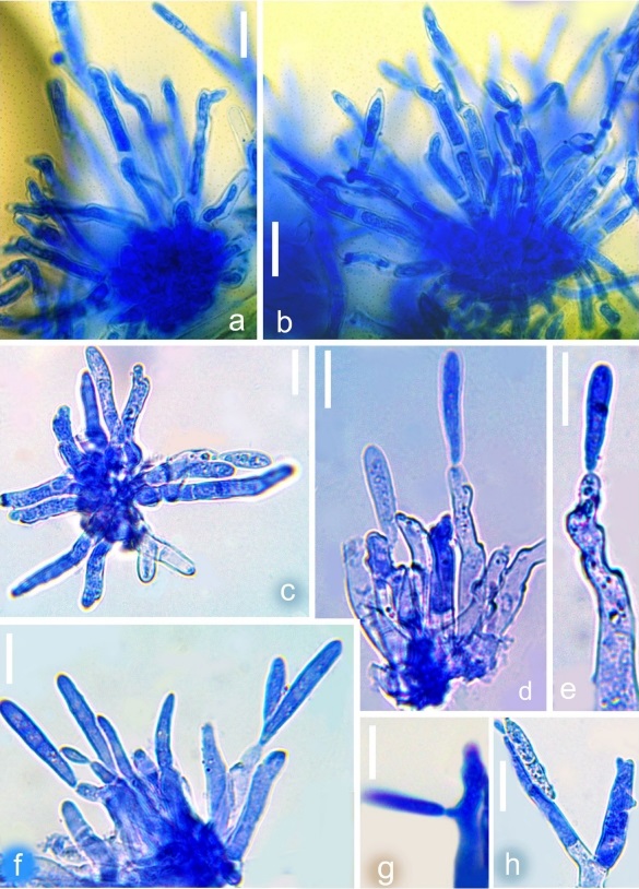
Figure 2 –Microphotographs of Neocercosporella peristrophes (AMH 10363). (a–c) Fascicles of conidiophores, (d–g) conidiophores with conidia, (h) branched conidiophores. Bars: 10 µm
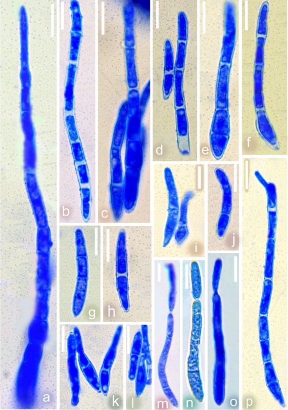
Figure 3 – Microphotographs of Neocercosporella peristrophes (AMH 10363). (a–l) Conidia, (m–o) catenate conidia, (p) germinating conidium. Bars: 10 µm.

Figure 4 –Scanning electron microphotographs of Neocercosporella peristrophes (AMH 10363). (a) Initial stage of development of conidiophores through stomata, (b, c) fascicles of conidiophores, (d) polyblastic conidiogenous cell (yellow arrows), (e–g) top view of conidiogenous loci, (h, i) lateral view of conidiogenous loci, (j, k) conidia, (l, m) Hila of conidia. Bars: (a–c)=10 µm, (d–i)=1 µm, (j, k)=10 µm, (l, m)=1 µm.