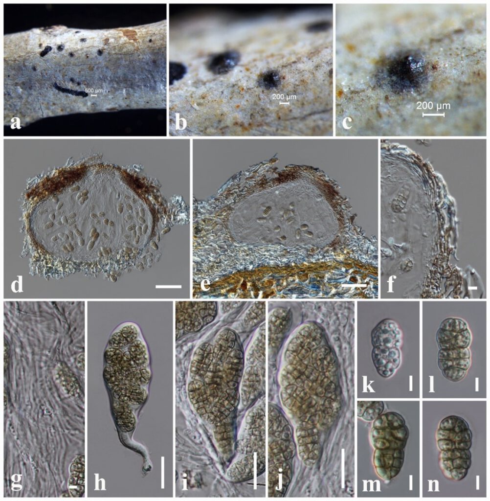
Muriformispora magnoliae N.I. de Silva, S. Lumyong & K.D. Hyde, sp. nov.,
MycoBank number: MB; Index Fungorum number: IF; Facesoffungi number: FoF 13098;
Etymology: Name reflects the host genus Magnolia, from which the new species was isolated.
Holotype: MFLU 18-2645.
Saprobic on dead twigs attached to Magnolia sp. Sexual morph: Ascomata 170–220 μm high × 200–260 μm diam. ( = 200 × 240 µm, n = 10), black, globose to subglobose, solitary, scattered, immersed to slightly erumpent, forming black spots on host surface, uni-loculate, ostiolate. Ostiole 50–70 μm wide, central. Peridium 14–28 μm, composed of several layers of hyaline, light brown to dark brown cells of textura angularis. Hamathecium composed of dense, 1.5–2 μm wide, filamentous, cellular pseudoparaphyses, with indistinct septa. Asci 80–95 × 25–30 μm ( = 85 × 28 μm, n = 20), 8-spored, bitunicate, fissitunicate, pyriform, pedicellate, with furcate to obtuse end, apically rounded. Ascospores 19–24 × 8–12 μm ( = 22 × 10 μm, n = 30), overlapping, 1-3-seriate, broadly ellipsoidal, muriform, 4–5 transverse septa and 2–3 longitudinal septa with apex rounded and basal end acute or truncate, at first hyaline becoming olivaceous-brown at maturity, constricted at the central septum and slightly at the other septa, with guttules in almost every cell, smooth-walled. Asexual morph: Not observed.
Culture characteristics – Colonies on PDA reaching 25 mm diameter after 1 week at 25 °C, colonies from above: olivaceous green, circular, flat, edge entire, margin well-defined, cottony to fairly fluffy with sparse aspects, white at the margin; reverse: grey from the centre of the colony, cream at margin.
Material examined – China, Yunnan Province, Xishuangbanna, dead twigs attached to the Magnolia sp. (Magnoliaceae), 27 March 2017, N. I. de Silva, NI261 (MFLU 18-2645, holotype), ex-type living culture, MFLUCC 19-0036.
GenBank numbers – (NI261); LSU: OL813499, SSU: OL824795, ITS: OM212459, tef1: ON303277, rpb2: ON502385, (NI261D); LSU: OL813500, SSU: OL824796, ITS: OM212460, tef1: ON303278.
Notes – The present phylogenetic analyses indicate that Muriformispora (Muriformispora magnoliae), constitutes a monophyletic clade distinctly separated from five genera, Brevicollum, Crassiparies, Medicopsis, Neohendersonia and Neomedicopsis in Neohendersoniaceae with 99% ML and 0.99 BYPP support (Fig. 19). Neohendersonia (Wijayawardene et al. 2016) and Neomedicopsis (Crous et al. 2019) are known from their asexual morph characteristics. Based on morphological characteristics of sexual morphs within species representing Neohendersoniaceae, the new genus (Muriformispora) is distinct from Brevicollum, Crassiparies and Medicopsis in having broadly ellipsoidal, muriform ascospores (de Gruyter et al. 2012, Tanaka et al. 2017). Muriformispora also has pyriform, pedicellate, apically rounded asci, with furcate to obtuse end, whereas Brevicollum, Crassiparies and Medicopsis have cylindrical or clavate asci (de Gruyter et al. 2012, Tanaka et al. 2017). In addition, ascomata structures of Brevicollum, Crassiparies and Medicopsis are different from Muriformispora. Medicopsis has stromata with poorly developed interior, immersed to erumpent from the bark with an ostiolar canal, circular to irregular in shape containing globose to subglobose, ostiolate, perithecia (de Gruyter et al. 2012). Brevicollum has scattered, sometimes 2–3 grouped, immersed, erumpent at ostiolar neck, globose to depressed globose ascomata (Tanaka et al. 2017). Crassiparies has scattered, immersed, erumpent at the ostiolar neck, subglobose, ostiolate ascomata (Tanaka et al. 2017). The new genus has black, globose to subglobose, uni-loculate, solitary, scattered, immersed to semi-immersed, ostiolate ascomata with black spots on the host surface. Based on morphological differences among other reported sexual morphs in Neohendersoniaceae and the phylogenetic analyses, we placed Muriformispora in the family Neohendersoniaceae. However, further collections are needed for the expansion of this genus.

Figure ** – Muriformispora magnoliae (MFLU 18-2645, holotype) a The specimen. b, c Appearance of ascomata on the host surface. d, e Vertical sections through ascoma. f Peridium. g Pseudoparaphyses. h–j Asci. k–n Ascospores. Scale bars: a = 500 μm, b, c = 200 μm, d, e = 50 μm, f, g, k–n = 5 μm, h–j = 20 μm.
