Morosphaeria velataspora (K. D. Hyde & Borse) Suetrong, Sakayaroj, E.B.G. Jones & Schoch, Stud. Mycol. 64:155 (2009). Fig. 110
≡ Massarina velatispora K.D. Hyde & Borse, Mycotaxon 27: 161 (1986).
MycoBank number: MB 542982; Index Fungorum number: IF 542982; Facesoffungi number: FoF 08305.
Saprobic on wood in mangrove habitats. Sexual morph: Ascomata 200–650 μm high, 350– 800 μm diam (x̅ = 371 × 522 µm, n = 5), immersed to erumpent solitary to gregarious, subglobose, raised, ostiolate, papillate, coriaceous, brown to black. Ostiole 65–330 μm long, 40–180 μm (x̅ = 196 × 108 µm, n = 5) diam., conical, black. Peridium 20–70 μm diam. (x̅ = 39 µm, n = 10), composed of thick-walled polyhedral cells of textura angularis fused with the host tissue. Hamathecium comprising 2–3 µm wide, numerous, septate, branched, filamentous to trabeculate pseudoparaphyses, resembling hyphae, embedded in a gelatinous matrix, anastomosing above the asci. Asci 180–260 × 20–40 μm (x̅ = 212 × 25 µm, n = 20), 8-spored, bitunicate, fissitunicate, clavate to cylindrical, short to long pedunculate, with an apical apparatus, thick-walled, bitunicate. Ascospores 45–55 × 12–17 μm, (x̅ = 48 × 14 µm, n = 50), obliquely 1-seriate, fusiform to ellipsoidal, hyaline, 1–3 septate, constricted at the septa, central cells larger, apical cells smaller and elongate, ascospores surrounded by a mucilaginous sheath. Asexual morph: Undetermined.
Culture characteristics – Ascospores germinating on 2 % sea water agar within 24 h with germ tubes produced from terminal ends. Colonies on malt extract sea water agar fast growth, white to pale pink, reverse pale brown, velvety, lobate, reaching 20 to 40 mm in diameter in 25 days at room temperature.
Material examined – India, Tamil Nadu, Parangipettai mangroves, (11.59°N 79.5°E), on decaying wood of Rhizophora mucronata (Rhizophoraceae), 23 April 2018, B. Devadatha, AMH- 9995, living culture, NFCCI-4425.
GenBank numbers – ITS: MK026766, LSU: MK026764, rpb-2: MN532683, SSU: MK026765, tef1: MN532688.
Notes – Morphology (Fig. 110) indicates that our new collection is identical to the species Morosphaeria velataspora. This result was supported by phylogenetic analyses in which our collection clustered with another strain of M. velatasporai with high bootstrap support (100 % MLBS, 1.0 PP, Fig. 42).
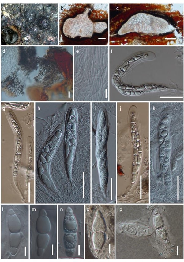
Figure 110 – Morosphaeria velatispora (NFCCI-4425). a Erumpent ascomata on decaying wood. b, c. Longitudinal sections of ascomata. d Peridial wall layers. e Hamathecium showing filamentous pseudoparaphyses. f–k Immature and mature asci. l–n Immature and mature ascospores. o–p Ascospores showing mucilaginous sheath in Indian ink. Scale bars: b, d, f–h = 50 μm, c, e, i–o = 10 μm.
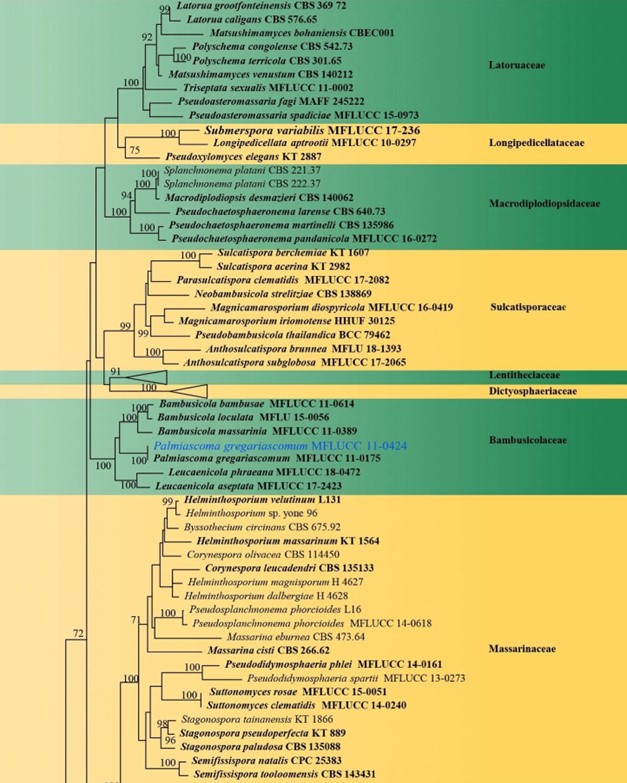
Figure 42 – Phylogram generated from maximum likelihood analysis (RAxML) of Pleosporales based on ITS, LSU, rpb-2, SSU and tef1 sequence data. Maximum likelihood bootstrap values equal or above 70 % are given at the nodes. An original isolate number is noted after the species name. The tree is rooted to Capnodium coffeae (CBS 147.52). The ex-type strains are indicated in bold.

Figure 42– Continued.
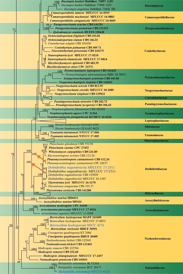
Figure 42– Continued.
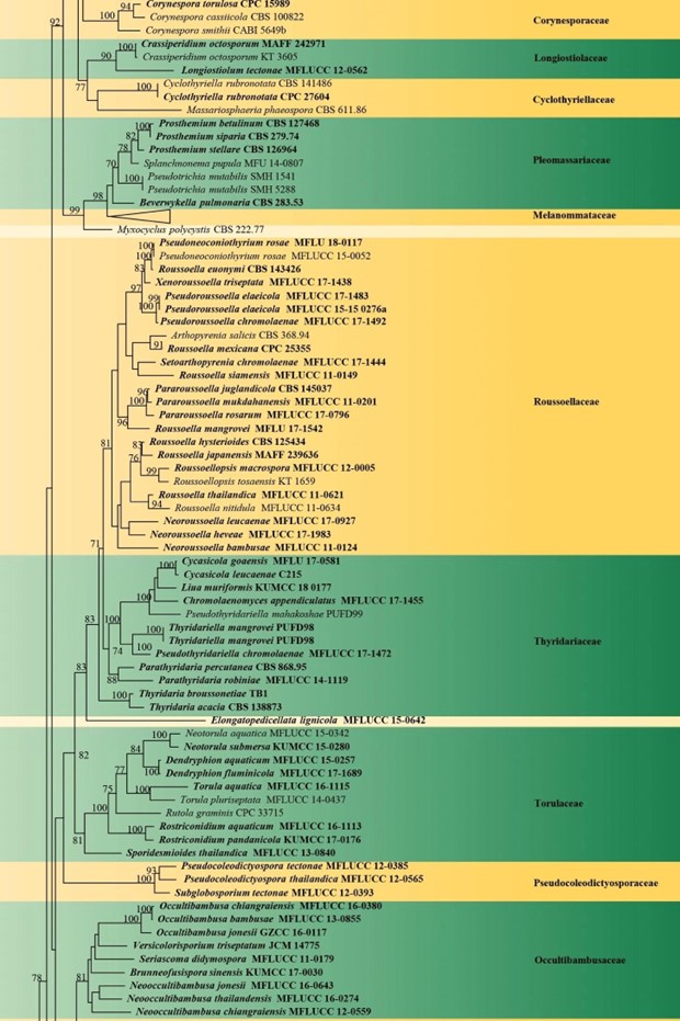
Figure 42– Continued.
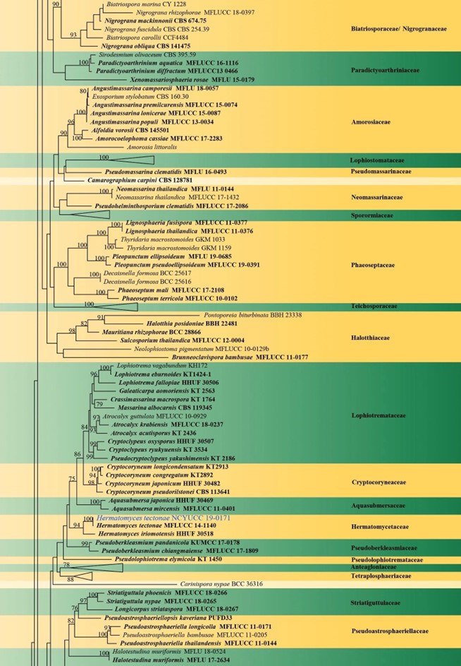
Figure 42– Continued.
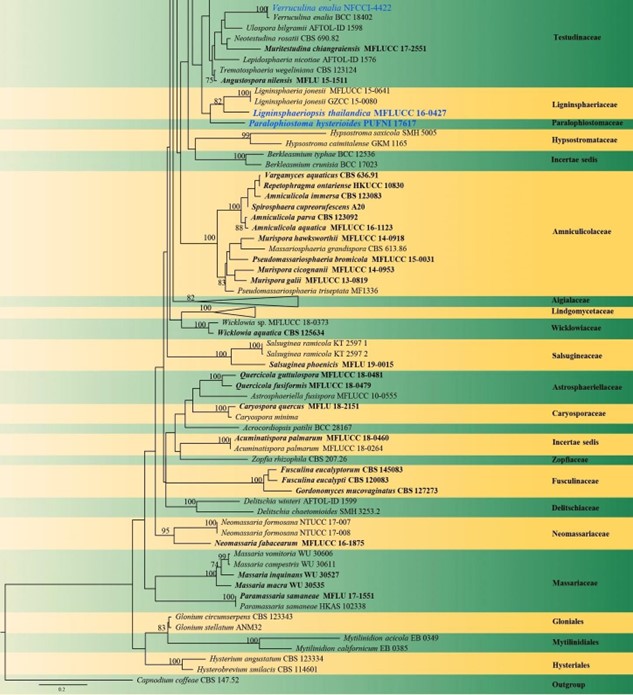
Figure 42– Continued.
