Microthyrium buxicola Hongsanan & K.D. Hyde.
Index Fungorum number: IF551429; Facesoffungi number: FoF00943; Fig. 2
Etymology – buxicola referring to the host on which the taxon was found.
Holotype – MFLU 15-0052
Epiphytic on the upper surface of dead fallen leaves, rarely on the lower surface, appearing as small black dots. Superficial hyphae absent. Sexual morph: Thyriothecia 190 – 240 μm diam. (x̄ = 215 μm, n = 5), superficial, mostly solitary, sometimes gregarious, light brown to brown, circular, flattened, basal peridium poorly developed, easily removed, with prominent, darker, central ostiole, pale brown and meandering and branched at the margin. Upper wall comprising brown cells of textura angularis, radiating in parallel lines from center to the outer rim. Pseudoparaphyses not seen. Asci 20 – 32 × 10 – 12 μm (x̄ = 30 × 11 μm, n = 10), 4 – spored, bitunicate, fissitunicate, fusiform to clavate, short pedicellate or apedicellate, apically rounded, with an ocular chamber. Ascospores 14 – 15 × 4 – 5 μm (x̄ = 14.4 × 4.6 μm, n = 10), 2 – 3 – seriate, fusiform to ellipsoidal, hyaline, 1-septate, not constricted at the septum, or slightly constricted at the septum when in 10 % lactic acid, apical cell slightly wider and longer than lower cell, smooth – walled, 2 – 3 – guttulate, with 3 – 6 – appendages near the apex, and 3 – 4 – appendages at the septum. Asexual morph: Undetermined.
Culture characteristics – Ascospores germinating on PDA at 25–30 °C in 12 h of light/12 h of dark, at first hyaline to brownish, becoming brown to reddish. Colonies slow growing reaching 1.5 cm diam. after 10 days, colony superficial to erumpent, very thin, surface smooth, difficult to remove, darker at the center.
Material examined – ITALY, on leaves of Buxus sp. (Buxaceae), 9 December 2014, E. Camporesi (MFLU 15- 0052, holotype); ibid., (KIB, isotype); ex-type living cultures MFLUCC 15-0212, MFLUCC 15-0213.
Notes – Microthyrium buxicola is most similar to M. nolinae A.W. Ramaley, but differs in lacking superficial hyphae (Ramaley 1999). Ascospores have up to 20 appendages around the upper cells in M. nolinae, while in M. buxicola there are 3 – 6 – appendages around the upper cell and 3–4-appendages at the septum. The species is also similar to M. guadalupensis A.W. Ramaley. Microthyrium guadalupensis has ascospores with 1 – 4 appendages at both ends, unlike in M. buxicola. Combined gene analysis of LSU and SSU sequences data indicate that M. buxicola groups in the Microthyriaceae clade sister to Micropeltidaceae clade and is a distinct species (Fig. 1).
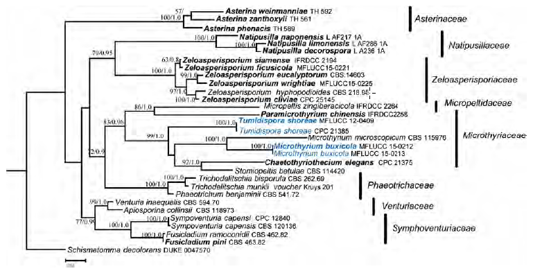
Fig. 1 Phylogram generated from maximum likelihood analysis based on combined LSU and SSU sequenced data. The first set of numbers above the nodes are maximum likelihood values above 50 % shown. The second set of numbers above the nodes are Bayesian posterior probabilities, with values above 0.9 shown. Strain numbers are indicated after species names. New sequence data are in blue bold, and ex-type strains are in black bold.
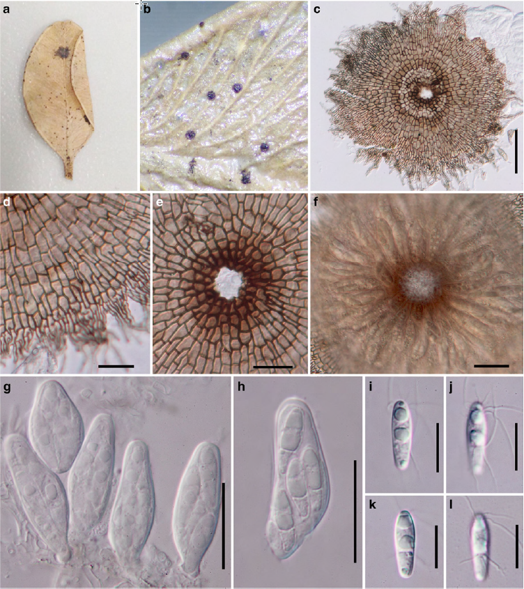
Fig. 2 Microthyrium buxicola (holotype). a Substrate b Thyriothecia on the surface of leaves c, f Thyriothecium when viewed in squash mounts and close-up of ascal arrangement d Upper wall when viewed in squash mount e Prominent darkened ring around the central ostiole g, h Asci with 4 – spores i, l Ascospores with appendages. Scale bars: c = 50 μm, d, i – l = 10 μm, e – h = 20 μm.