Melomastia fusispora W.L. Li, Maharachch. & Jian K. Liu, sp. nov
MycoBank number: MB 841499; Index Fungorum number: IF 841499; Facesoffungi number: FoF 10533;
Etymology: Refers to the shape of the ascospore.
Holotype: HKAS 121316
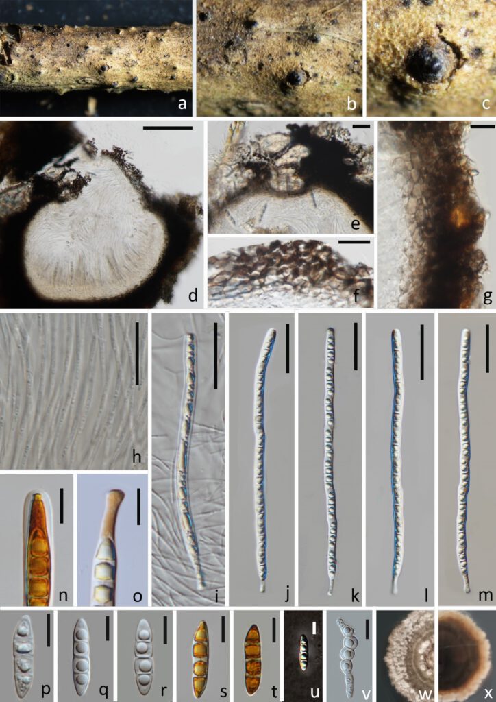
Saprobic on dead branches of Olea europaea. Sexual morph: Ascomata 432–624 × 527–618 μm diam. ( = 528 × 572 μm, n = 15), visible as numerous, black, cone-like structures on host surface, solitary, gregarious, immersed to erumpent through host tissue, globose to compressed globose, coriaceous to carbonaceous, dark brown to black, rough-walled, ostioles. Ostioles 19.14–343.5 × 65.8–283.5 μm ( = 174.5 × 181 μm, n = 15), central, carbonaceous, dark brown to black, papillate, periphyses filling the ostiolar canal. Peridium 25.5–61.5 μm ( = 41 μm, n = 15) wide, two-layered, an outer layer of cells of textura intricata composed of host cells interspersed with fungal hyphae and an inner layer of thick-walled cells of textura angularis. Hamathecium comprises numerous, 2–2.6 μm ( = 2.3 μm, n = 30) wide, dense, filiform, unbranched, aseptate pseudoparaphyses. Asci 200–231 × 7.6–9.2 μm ( = 215 × 8.4 μm, n = 30), 8-spored, bitunicate, cylindrical, slightly flexuous, apically round, with well-developed ocular chamber, cylindrical pedicellate 11–17.5 × 3.5–4.3 μm ( = 14 × 3.9 μm, n = 30). Ascospores 27.5–32 × 6.5–7.5 μm ( = 30 × 7 μm, n = 30), uniseriate, partially overlapping, hyaline, fusiform, with rounded to acute ends, narrow towards apex, 3-septate, constricted at the central septum, with guttules in each cell, surrounded by an irregular and thin gelatinous sheath. Asexual morph: Undetermined.
Culture characteristics: Colonies on PDA reaching 40 mm diam. after 4 weeks at 25 °C. Culture from above, brownish grey, forming zonate grey, fluffy mycelium at the edge; reverse dark brown, greyish orange at the edge.
Material examined: China, Sichuan Province, Chengdu City, Shuangliu District, Olive Base, 30°33.25′N, 103°99.62′E, at an altitude of 438 m (mountainside), on dead branch of Olea europaea, 27 March 2021, W.L. Li, T. Zhang, GL 139 (HKAS 121316, holotype; HUEST 21.0002, isotype), ex-type living culture CGMCC3.20618 = UESTCC 21.0002; ibid., GL 136 (HUEST 21.0001, paratype), living culture UESTCC 21.0001.
GenBank numbers –LSU, ITS, SSU, TEF, RPB2
Notes: Phylogenetic analysis showed that Melomastia fusispora is sister to M. winteri with high support (97% ML BS, 100% MP BS, 1.00 PP). Melomastia fusispora resembles M. winteri in having cylindrical asci and fusiform, 3-septate ascospores. However, M. fusispora has longer asci (200–231 vs. 165–189 μm), longer pedicellate (11–17.5 vs. 4.8–6.5 μm) and longer ascospore (27.5–32 vs. 25–30 μm) when compare to M. winteri. In addition, the ascospores ends of the Melomastia winteri are usually pointed, which is distinguished from M. fusispora ascospores which has round to acute ends.

Figure 1. Melomastia fusispora (HKAS 121316, holotype). (a–c) Ascomata on the substrate. (d) Vertical section of the ascomata. (e) Vertical section of the ostiole. (f, g) Peridium (h) Paraphyses. (i–m) Asci. (n) Ocular chamber in Lugol’s lodine. (o) Pedicel in Lugol’s lodine. (p–u) Ascospora. (s,t in Lugol’s lodine, u in India Ink). (v) Geminating ascospore. (w) Upper view of the colony after 14d. (x) Reverse view of the colony after 14d. Scale bars: d = 100 μm, e, g = 20 μm, f, n–u = 10 μm, h–m = 40 μm.
