Melanodevriesia melanelixiae H.L. Si, W.Q. Cao, T. Bose, sp. nov.
MycoBank number: MB 840429; Index Fungorum number: IF 840429; Facesoffungi number: FoF 11078; Figs 2, 3.
Etymology: The name is derived from the lichen Melanelixia subargentifera, from which both isolates of this fungus were obtained.
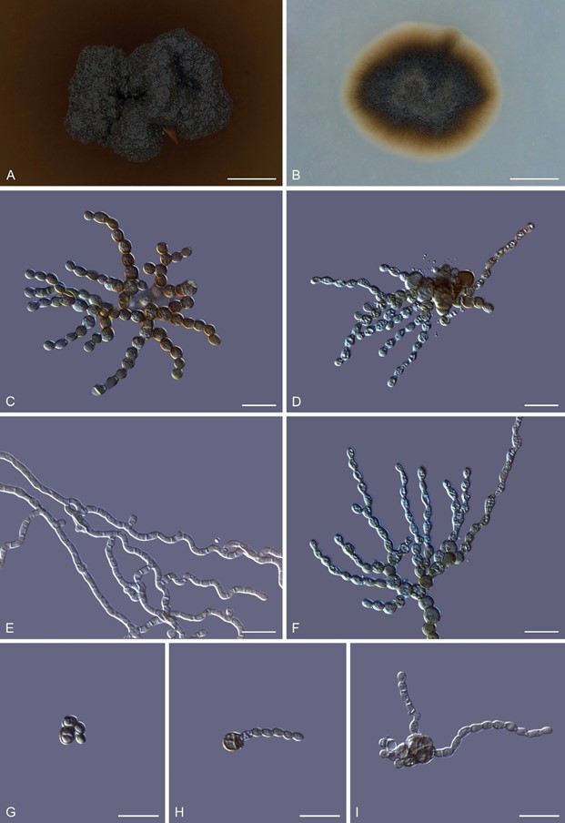
This fungus can exist in both a yeast-like and a mycelial state. The yeast-like thallus produces pseudohyphae through budding. These pseudohyphae are branched, septate, constricted at the septa, composed of oval to urceiform cells, hyaline to brown in colour, smooth-walled, guttulate, measuring 1.4–3 × 2.3–4.6 μm (Fig. 2). In the mycelial state, hyphae grow into the substrate. Hyphae branched, septate, smooth-walled, smooth or corrugated, cylindrical, hyaline to pale brown in colour, measuring 1.3–2 μm wide (Fig. 2). Chlamydospores spherical to ovoid in shape, solitary often monilioid forming radiating clusters, smooth-walled, pale brown to dark brown in colour, usually aseptate, rarely septate, guttulate, measuring 2.8–4.2 × 2.8–4.8 μm (Fig. 2). Chlamydospores geminate into yeast-like unicellular conidia that are globose to sub- globose in shape, pale brown to dark brown in colour, thick-walled, measuring 4–7.3 × 3.6–6.2 μm (Fig. 2). These unicellular cells multiply through budding (Fig. 2) forming multicellular structures from which pseudohyphae emerge randomly (Fig. 2). No sexual reproductive structures were observed.
Culture characteristics: After 12 wk on PDA, the surfaces of the colonies were dark brown to black with the reverse dull brown in colour, erumpent, hollow, with irregular margins, rarely with a few aerial mycelia. After a few weeks after subculturing, the colony stains the PDA brown. Colonies slow-growing, reaching 3.1 ± 0.1mm diam after incubating at 25 °C for 12 wk (Fig. 2). After 8 wk on OA, the colonies are round to oval in shape, with smooth margins, surface taupe brown to olive-brown with the reverse taupe brown in colour. Colonies are slow-growing on OA yet faster than on PDA, reaching 5.42 ± 0.2 mm diam after incubating at 25 °C for 8 wk (Fig. 2).
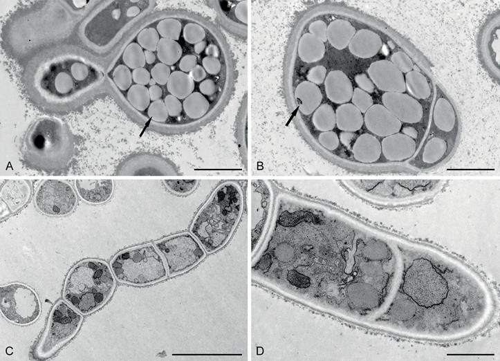
Intracellular oil bodies: The TEM of yeast-like cells grown on PDA revealed thick cell walls with many inconspicuous oil bodies concealing the other cell organelles. Hyphae grown on OA lacked thick cell walls and intracellular oil bodies (Fig. 3).
Typus: China, Inner Mongolia Autonomous Region, Chifeng, Balin Right Banner, Mt. Qingyangcheng, 44°13’46″N, 118°44’57″E, 1 498.8 m alt, isolated from the medullary tissue of Melanelixia subargentifera, 7 Jul. 2019, H.L. Si (holotype HMAS 350275; ex-type culture CGMCC3.20308).
Notes: Melanodevriesia melanelixiae differs from X. strelitziicola in that it contains at least two thallus morphologies and chlamydospores. Besides this, we did not observe any sexual reproductive structures (Crous et al. 2009, 2019).

Fig. 2. Morphology of Melanodevriesia melanelixiae sp. nov. (ex-type CGMCC3.20309). Colony morphology on potato dextrose agar (A) and oatmeal agar (B). C, D. Microscopic structures of 14-d-old culture growing on PDA medium with yeast-like unicellular morph forming pseudohyphae through budding. E. Straight and corrugated septate hyphae produced by the mycelial state of the fungus. F. A cluster of monilioid chlamydospores. G–I. Single chlamydospores germinating into unicellular cells that multiply through budding, forming a multicellular structure from which pseudohyphae emerge. Scale bars: A, B = 2 mm; C–I = 10 μm.

Fig. 3. Transmission electron microscopic images of pseudohyphae and mycelium of Melanodevriesia melanelixiae sp. nov. (ex-type CGMCC3.20309).
A, B. Budding yeast-like unicellular cell with thick cell walls. Multiple intracellular oil bodies concealing the cell organelles (indicated with arrows). C,
D. Septate hyphae with a thin cell wall that is devoid of intracellular oil bodies. Due to the lack of intracellular oil bodies, various cell organelles are visible. Scale bars: A, B = 2 μm; C = 10 μm; D = 1 μm.
