Lophiostoma caulium (Fr.) Ces. & De Not., Schem. di Classif. Sferiacei: 45 (1863)
Facesoffungi number: FoF00881; Fig. 2.
Saprobic on Salvia. Sexual morph: Ascomata 190 – 210 μm high × 122 – 190 μm diam. (x̄ = 198 × 171 μm, n = 5), scattered to gregarious, semi-immersed to erumpent, dark brown to black, globose to subglobose, ostiolate. Ostiole 80 – 90 μmhigh, 83 – 110 μmdiam. (x̄ = 84×95 μm, n=5), slitlike, with a small, to large, flat, crest-like apex, which is variable in shape; apex composed of pseudoparenchymatous cells. Peridium 19 – 23 μm thick at the sides, broad at the apex, thinner at the base, composed of 4 – 5 layers of brown to dark brown cells, arranged in a textura angularis, fusing at the outside with the host cells. Hamathecium of 1.5 – 2.5 μm diam., septate, long, anastomosing and branched, cellular pseudoparaphyses, embedded in gelatinous matrix. Asci 64 – 91 × 8 – 10 μm (x̄ = 82 × 9 μm, n = 30), 8 – spored, bitunicate, fissitunicate, cylindrical to clavate, with furcate pedicel, rounded at the apex, with an ocular chamber. Ascospores 20 – 24 × 4 – 6 μm (x̄ = 22 × 5 μm, n = 30), uniseriate or overlapping biseriate, pale brown to dark brown, narrowly fusiform with acute ends, 3 – 5 – septate, slightly constricted at each septum, with a distinct guttule in each cell, smooth – walled, with mucilaginous appendages, drawn out at the ends. Asexual morph: Coelomycetous. Conidiomata superficial, dark brown to black, globose, uniloculate, solitary to scattered. Pycnidial wall thickwalled, multi-layered, with inner layer comprising several cell-layers, comprising brown-walled cells of textura angularis. No spores were produced.
Culture characteristics – Ascospores germinating on MEA within 36 h. Colonies growing on MEA, rather slow growing, reaching 0.2 mm diam. in 1 week at 28 °C. Mycelium superficial, felty, gummy, grey.
Material examined – ITALY, Forlì-Cesena Province, Teodorano di Meldola, dead stem of Salvia sp., 13 October 2013, E. Camporesi (MFLU 15-1074, reference specimen designate here); ex-type living culture, MFLUCC 15-0176, BIOTEC.
Notes – Our material of Lophiostoma caulium (Fr.) Ces. de Not. fits well with the description provided by Cesati and De Notaris (1863). In our collection of L. caulium, the ascomata are slightly smaller than that in the type, but the size of peridium and ascospores are in the range given. The ascomata, asci and ascospores of our reference specimen provided here are also typical of the L. caulium. Our specimen of L. caulium was collected from a different host than that of the type, thus we propose this as reference specimens until collections from the same host and location can be obtained. In our phylogeny Lophiostoma caulium (MFLUCC15-0176) forms a well-supported (BS 100) clade sister to Lophiostoma caulium “var. a” (JCM 17669) in the family Lophiostomataceae (Fig. 1).
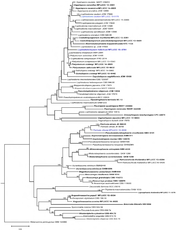
Fig. 1 Phylogram generated from maximum likelihood analysis based on combined LSU, ITS, EF and SSU sequenced data for the family Lophiostomataceae. Maximum likelihood bootstrap support values greater than 50 % are above the nodes. The ex-type strains are in bold and the new isolates are in blue. The tree is rooted to Melanomma pulvispyrius (strain CBS 124080).
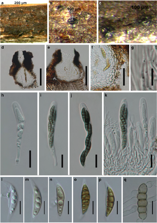
Fig. 2 Lophiostoma caulium (reference specimen) a – c Appearance of immersed ascomata on host surface d, e Hand sections of ascomata. Note the ostiole, with a small to large, flat, crest-like apex f Peridium g. Hamathecium h – k Asci with ascospores l – p Ascospores o. Germinating ascospore. Scale bars: d, e = 100 μm, f, h – k = 20 μm, g = 10 μm, l – q = 10 μm.