Lauriomyces synnematicus Somrithipol, sp. nov.
Index Fungorum number: IF551016, Facesoffungi number: FoF 00554; Figs. 1, 2
Etymology: synnematicum, refers to the synnematal structure
Holotype: SFC 2156 in BBH
Saprobic on decaying leaves. Sexual morph Undetermined. Asexual morph Colonies scattered, white conidial sporulation on scattered synnemata. Synnemata solitary or in groups, scattered, black, 50 – 225 μm high, 10 – 40 μm diam. with 5 – 20 filaments closely adpressed along most of their length. Filaments septate, thick and smooth-walled, brown to dark brown, 5 – 6 μm wide, with terminal branches. Branches cylindrical, thin and smooth-walled, hyaline to subhyaline. Primary branches in clusters of 3 – 5 at the apex of the filament, 6 – 7.5 μm long (x̄ = 7 ± 0.68 μm, n = 5), 1 – 2.5 μm wide (x̄ = 2 ± 0.28 μm, n = 5). Subsequent branches in clusters of 3 – 5, 6 – 7.5 μm long (x̄ = 6.75 ± 0.68 μm, n = 5), 1 – 2 μm wide (x̄ = 1.4 ± 0.28 μm, n = 5). Ramoconidia and conidia holoblastic, cylindrical, one-celled, hyaline to subhyaline, thinand smooth-walled, in acropetal branched chains, 4.5 – 7.5 μm long (x̄ = 5.4 ± 0.61 μm, n = 50), 0.7 – 1.3 μm wide (x̄ = 1 ± 0.12 μm, n = 50).
Material examined – THAILAND, Nakhon Rachasima, on decaying leaves, 23 August 2008, B. Thongnuch (SFC 2156 in BBH, holotype).
Notes – Lauriomyces synnematicus differs from other species of the genus in possessing synnemata. The synnema is an important characteristic to distinguish several hyphomycete species, for example: Janetia synnematosa (Sivanesan and Hsieh 1990), Melanographium selenioides (Ellis 1963), Memnoniella stilboidea (Ellis 1976) and Dictyoarthrinium synnematicum (Somrithipol 2007). Conidia of L. synnematicum are rather narrower than those of the other species in the genus with cylindrical conidia. Somrithipol and Jones (2007) illustrated the gelatinization process during the conidial formation of L. cylindricus, resulting in deposition of mucilage between the conidia to join them into a persistent chain. This conidial chain is the only character distinguishing the genus Lauriomyces from Haplographium. In L. synnematicum, the mucilage was less evident but some persistent chains could be observed.

Fig. 1 Lauriomyces synnematicus (holotype). a A synnema with conidial mass b Synnematous filaments bearing conidiogenous cells and branched chains of conidia and c Cylindrical conidia. Scale bars: a = 50 μm, b, c = 10 μm.
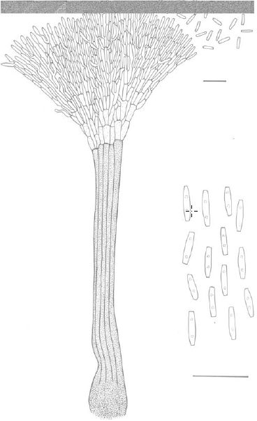
Fig. 2 Lauriomyces synnematicus (holotype). Line drawing of a synnema with filaments bearing branched chains of conidia and cylindrical conidia. Scale bars: = 10 μm.