Kalmusia spartii Wanasinghe, Camporesi, E.B.G.Jones & K.D. Hyde.
Index Fungorum Number: IF550895, Facesoffungi number: FoF00385; Figs. 2 and 3
Etymology – Named after the host genus from which it was collected, Spartium.
Holotype – MFLU 14–0751
Saprobic on dead herbaceous branches. Sexual morph Ascomata 350 – 450 μm high × 250–400 μm diam. (x̄ = 395.3 × 323.7 μm, n = 10), solitary, scattered, immersed to erumpent, globose or subglobose, coriaceous, wall black,with orwithout papilla, ostiolate. Ostiole 150 –200 μm high, 90 – 130 μm diam. (x̄ = 174.3 × 109 μm, n = 10), blackish-brown, smooth, ostiolar canal filled with hyaline cells. Peridium 15 – 30 μm wide at the base, 25 – 50 μm wide at the sides, thick, 1-layered, composed of small heavily pigmented thick-walled cells of textura angularis. Hamathecium comprising numerous, 1.9μm (n = 30) wide, filamentous, branched septate, pseudoparaphyses. Asci 110 – 130 × 10 – 16 μm (x̄ = 119.6 × 13.9 μm, n = 40), 8- spored, bitunicate, fissitunicate, clavate, with a long, narrow, furcate pedicel, up to 30–35 μm long, with a minute ocular chamber. Ascospores 19 – 22 × 5.5 – 9 μm (x̄ = 20.2 × 7.6 μm, n = 50), overlapping 1 – 2-seriate, narrowly ovoid to clavate, 3- distoseptate, constricted at the septa, initially hyaline, becoming yellowish-brown at maturity, narrowly rounded at both ends, smooth-walled, not surrounded by a mucilaginous sheath. Asexual morph Conidiomata 300 – 550 μm diam., 200 – 250 μm high, superficial or immersed in the agar, dark brown to black, clothed with white hyphal projections, simple cavities. Conidiomatal wall composed of a 40 – 50 μm wide outer layer and 10 – 20 μm wide inner layer of cells of textura angularis. Conidiogenous cells 5 – 8 × 2 – 4 μm discrete, assembled into protruding masses, or integrated in very compact conidiophores. Conidia 2.5 4 × 1.5 – 2.5 μm (x̄ = 3.2 × 1.9 μm, n = 20) narrowly ellipsoidal or short-cylindrical, straight or slightly curved, rounded at both ends, one-celled, with one or two small, polar guttules, initially hyaline, becoming light brown, smooth-walled.
Culture characters – Colonies on PDA reaching a diam. of 30 – 35 mm in 21 d, flat, with undulate to lobate margin, hyaline, covered by thin, felty, white, aerial mycelium, honeyyellow in reverse, sporulation after 8 weeks.
Material examined – ITALY, Forlì-Cesena Province: Castello di Corniolo, Santa Sofia, dead and hanging branches of Spartium junceum L. (Fabaceae), 15 March 2013, E.Camporesi (MFLU 14 – 0751, holotype); ex-type living culture, MFLUCC 14 – 0560. GenBank ITS: KP744441; LSU: KP744487; SSU: KP753953.
Notes – Multi-gene phylogenetic analyses indicated that Kalmusia spartii belongs to Didymosphaeriaceae and grouped together with other Kalmusia strains. Kalmusia spartii has a similar morphology to K. ebuli, but in our phylogenetic tree, K. ebuli and K. spartii were well resolved (Fig. 1).
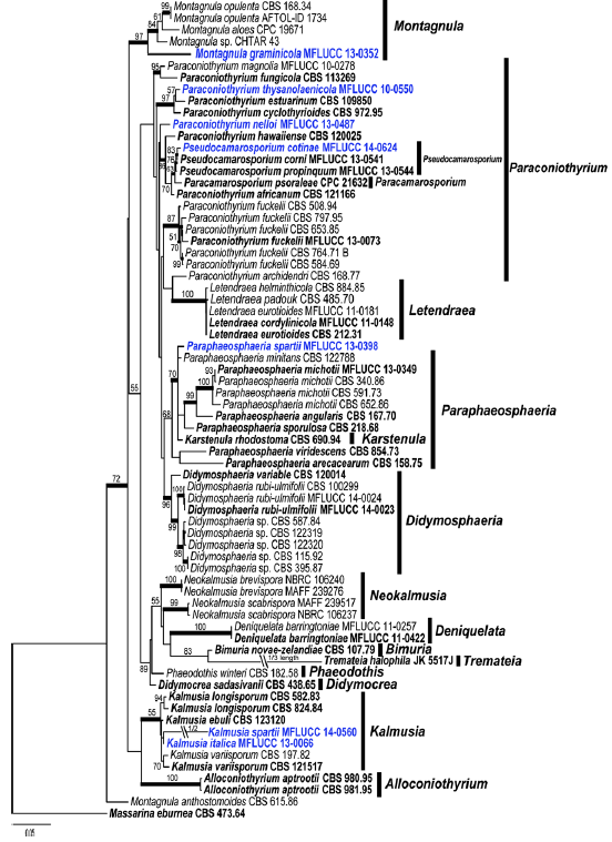
Fig. 1 Phylogram generated from Maximum likelihood analysis based on combined SSU, LSU, ß – tubulin and ITS sequence data of Didymosphaeriaceae. Maximum likelihood bootstrap support values greater than 50 % are indicated above and below the nodes, and branches with Bayesian posterior probabilities greater than 0.95 are given in bold. The ex – types (reference strains) are in bold; the new isolates are in blue. The tree is rooted with Massarina eburnea CBS 473.64.
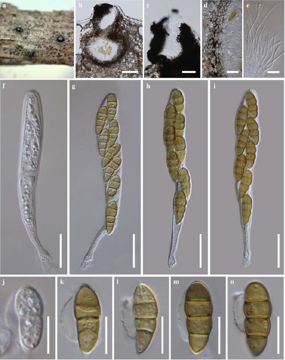
Fig. 2 Kalmusia spartii (holotype) a Ascomata on host substrate b Section of ascoma c Close up of ostiole d Peridium e Pseudoparaphyses f – i Asci j – n Ascospores. Scale bars: b = 100 μm, c = 50 μm, d – i = 20 μm, j – n = 10 μm.
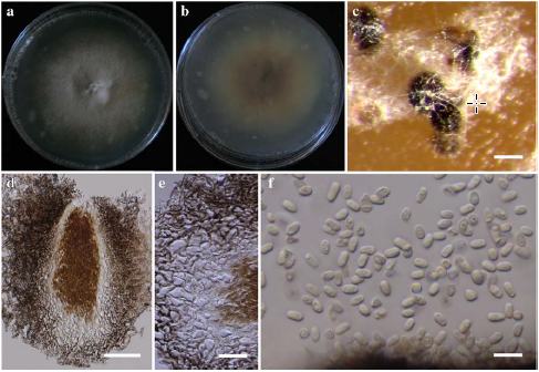
Fig. 3 Asexual morph of Kalmusia spartii (ex-type culture) a, b Colonies on PDA (b from below) c conidiomata d Longitudinal sections of conidiomata e Conidiomatal wall f Conidia. Scale bars: c = 100 μm, d = 50 μm, e = 20 μm, f = 5 μm.