Jahnula dianchia S.K. Huang & K.D. Hyde, Mycol. Progr. 17: 549 (2018).
MycoBank number: MB 553200;Index Fungorum number: IF 553200; Facesoffungi number: FoF 03149; Fig. 36
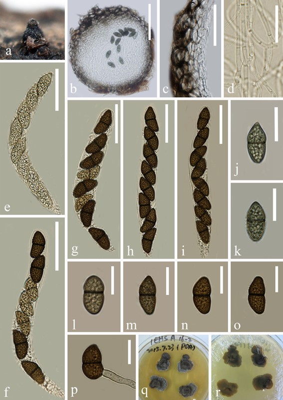
Saprobic on decaying wood submerged in freshwater habitats. Sexual morph: Ascomata 285–390 μm high, 250–350 μm diam, perithecial, solitary, superficial to semi-immersed, obpyriform to subglobose, black, papillate, ostiolate. Peridium around 40 μm thick, membranous, composed of brown to hyaline cells of textura angularis. Hamathecium comprising 2–3 μm wide, septate, branched, filiform, cellular pseudoparaphyses, embedded in a gelatinous matrix. Asci 150–170 µm ( x̄ = 160 µm, SD = 10, n = 15) long, 15–17 μm ( x̄ = 16 µm, SD = 1, n = 15) wide, 8-spored, bitunicate, fissitunicate, cylindrical, pedicellate, rounded at apex, with a distinct ocular chamber. Ascospores 27–29 µm ( x̄ = 28 µm, SD = 1, n = 20) long, 11–13 μm ( x̄ = 12 µm, SD = 1, n = 20) wide, uni-seriate, oval to broadly ellipsoid, slightly curved, initially pale brown, becoming dark brown at maturity, 1-septate, verruculose, multiguttulate. Asexual morph: Undetermined.
Material examined: China, Yunnan Province, saprobic on decaying wood submerged in Erhai Lake, December 2014,
Z.L. Luo, S-364 (HKAS 92632), living culture MFLUCC 16-0983; saprobic on decaying wood submerged in Erhai Lake, December 2014, H.Y. Su, S-460, living culture MFLUCC 16-1353; saprobic on decaying wood submerged in a freshwater stream in Cangshan Mountain, June 2016, S.M. Tang, S-740.
GenBank numbers: ITS: MH793537, LSU: MH793543 (MFLUCC 16–0983); ITS: MH793538, LSU: MH793544 (MFLUCC 16-1353), LSU: MT797171 (S-740).
Notes: Jahnula dianchia was collected from a freshwater lake in Yunnan Province, China. During our study of lignicolous freshwater fungi from Northwestern Yunnan Province, three isolates were obtained from different collections. The morphology of those isolates fit well with Jahnula dianchia, and the phylogenetic analysis showed that our isolates cluster with this species with strong support. We therefore identify our isolates as J. dianchia based on morphology and phylogeny.

Fig. 36 Jahnula dianchia (HKAS 92632). a Appearance of ascomata on substrate. b Vertical section of ascoma. c Structure of peridium. d Pseudoparaphyses. e–i Asci. j–o Ascospores. p Germinating ascospore. q, r Colony on MEA. Scale bars: b = 150 μm, c, e–i = 50 μm, j–p = 20 μm, d = 15 μm
