Hypoxylon lenormandii Berk. & M.A. Curtis [as ‘lenormandi’], in Berkeley, J. Linn. Soc., Bot. 10(no. 46): 385 (1868) [1869]
Index Fungorum number: IF 152747; Facesoffungi number: FoF 00300, Fig. 1
Holotype – K 42245
Saprobic on decorticated wood. Sexual morph: Ascostromata 0.1–1 × 0.04–0.1 cm diam. (x̄ = 0.6 × 0.08 cm), glomerate and effuse-pulvinate, stromata almost single and rosellinoid, connected by very thin stromatal tissue at the base, with very conspicuous perithecial mounds, half or sometimes entire perithecial mounds exposed, surface greyish-sepia (106), fuscous (103), or brown-vinaceous (84), dull orange brown to dark brown granules immediately beneath surface and between perithecia, with KOH extractable pigments hazel (88), fulvous (43), umber (9) or ochreous (44), the tissue below the perithecial layer inconspicuous, perithecia 0.3–0.5 mm diam. (x̄ = 0.4 mm), immersed, sphaerical, ostioles slightly papillate, without apparent disk formation. Paraphyses not seen. Asci (120–)122–150(–153) × (5.3–)5.5–8.5(–8.7) μm (x̄ = 134 × 7.1 μm), 8-spored, unitunicate, cylindrical, pedicellate, with apical ring not bluing in Melzer’s reagent, discoid, 0.2–0.5 × 2–3 μm broad (x̄ = 0.3 × 2.7 μm). Ascospores (10.3–)10.5–15(–15.4) × 6–8.5 μm (x̄ = 13.8 × 7.6 μm), uniseriate, one-celled, ellipsoid-inequilateral, with narrowly rounded ends, brown to dark brown, with straight, to slightly sigmoid germ slit more or less entire spore-length, perispore dehiscent in 10 % KOH, epispore smooth. Asexual morph: Conidiogenous structures nodulisporium-like, hyaline. Conidiogenous cells 12–17 × 3–3.5 μm (x̄ = 15 × 3.3 μm), hyaline, smooth. Conidia 5.5–7 × 2.5–3 μm (x̄ = 6.2 × 2.7 μm), hyaline, ellipsoid.
Culture characters – Colonies on OA at 25–28 °C reaching the edge of 4cm in 7 days, at first white, becoming hazel (88) to greyish-sepia (106), azonate with diffuse margins, reverse at first fuscous (103) and turning black after 10–12 days. Sporulating regions scattered over the surface of colony particularly at the centre, hazel (88).
Material examined – CUBA, on bark, C. Wright 485 (K42245, holotype); THAILAND, Chiang Mai, Doi Suthep, on decaying wood, 10 November 2012, D.A. Daranagama and K.D. Hyde AXL 104 (MFLU 12–0831), living cultures, MFLUCC 13–0311; ibid., Chiang Rai, Doi Mae Saloung, on dead bamboo clumps, 12 December 2012, D.A. Daranagama and K.D. Hyde AXL 112 (MFLU 12–0843), living culture, MFLUCC 13–0304; ibid., Mae Fah Luang University, on decaying wood, 20 December 2012, D.A. Daranagama and K.D. Hyde AXL 115 (MFLU 13–0847), living cultures, MFLUCC 13–0120.
GenBank accession numbers – ITS: KM039135; LSU:KM039136; RPB2: KM039137.
Notes – Hypoxylon lenormandii, with its glomerate stromata can easily be distinguished from other species, such as H. undulatum. Both taxa have very conspicuous stromata, but only H. lenormandii has characteristic glomerate stromata, more similar to the Rosellinoid type, which in fact led mycologists to name this fungus several times under Rosellinia. Hypoxylon undulatum is characterized by the lack of KOH extractable pigments (Ju and Rogers 1996). Therefore, our specimen cannot be placed under H. undulatum. Hypoxylon sublenormandii described by Suwannasai et al. (2005) from Thailand is similar to H. lenormandii. However, the stromatal surface of the new taxon is strongly reddish brown, while H. lenormandii is greyish sepia, fuscous or brownvinaceous. In addition, asci (95–110×3.8–5μm) and ascospores of H. lenormandii are longer than H. sublenormandii (Ju and Rogers 1996, Suwannasai et al. 2005). Hypoxylon sublenormandii is so far known only from bamboo, whereas this specimen is from angiosperm wood. According to Ju and Rogers (1996), H. lenormandii can occur both on dicotyledonous and monocotyledonous substrates, indicating a fairly wide host range. According to Ju and Rogers (1996) the details in the description of H. lenormandii are compatible with this specimen and its nodulisporium-like asexual morph. We provide new ITS, LSU, RPB2 and β-tubulin gene sequence data from an authenticate strain of H. lenormandii which can be used in the future molecular analysis.
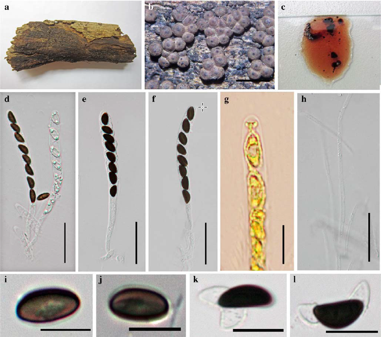
Fig. 1 Hypoxylon lenormandii (MFLU 12–0831) a Stromatal habit on wood b Perforations and ostioles seen fromabove c Stroma in side view d Cross section of the stroma showing perithecia e Perithecia from above f Formation of pigments in KOH g Mature ascus in water i Ascus in Melzer’s reagent showing non amyloid ascal apical apparatus j Ascospore in water k Dehiscent perispore in KOH l Ascospore showing germ slit. Scale bars: a-c=5 mm, d, f=30μm, g-l=10μm.