Hydnophlebia fissurata Authority.
MycoBank number: MB 843313; Index Fungorum number: IF 843313; Facesoffungi number: FoF 12683;
Description
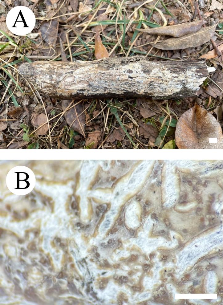
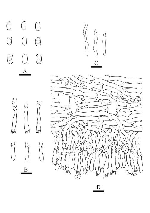
Basidiomata annual, resupinate, ceraceous, without odor or taste, when fresh, becoming hard upon drying, up to 9 cm long, 2 cm wide, 300–600 µm thick. Hymenophore grandinoid, cream to buff when fresh, buff to pale brown upon drying, cracking. Sterile margin narrow, cream, minutely fibrillose. Hyphal structure monomitic; generative hyphae simple-septate, colorless, thin-walled, IKI–, CB–; tissues unchanged in KOH. Subicular hyphae unbranched, 4.5–6.5 μm in diameter; subhymenial hyphae unbranched, 2–4 μm in diameter; the presence of numerous yellow to yellowish brown gelatinous substances among generative hyphae. Hymenium cystidia absent; cystidioles colorless, thin-walled, 13.6–28 × 1.3–3.6 µm; basidia cylindrical, with four sterigmata and a basal simple-septate, 21–35 × 2.8–5 µm. Basidiospores ellipsoid, colorless, thin-walled, smooth, often with 1–2 oil drops, IKI–, CB–, (2.8–)3–3.8 × 1.6–2.3 µm, L = 3.23 µm, W = 1.93 µm, Q = 1.72 (n = 30/1).
Material examined: China, Yunnan Province, Kunming, Xishan District, Haikou Forestry Park, E 103°03′, N 25°37′, alt. 2150 m, on the fallen branch of angiosperm, 16 Septemper 2017, C.L. Zhao, CLZhao 2900 (SWFC).
Distribution: The species is known from Yunnan Province, China, in a subtropical evergreen broad-leaved forest. It grows on moderately decayed angiosperm wood and causes white rot.
Sequence data: ITS: MW732402 (ITS5/ITS4); LSU: MW724794 (LROR/LR7); RPB1: ON892527 (RPB1-Af/RPB1-Cr); EF1a: ON968926 (EF1-983F/EF1-2218R); RPB2: ON892536 (bRPB2-6F/bRPB2-7.1R); mtSSU: MW732762 (MS1/MS2)
Notes: Hydnophlebia fissurata groups with Phlebia acanthocystis Gilb. & Nakasone and P. caspica Hallenb., however, morphologically P. acanthocystis differs in its odontoid to hydnoid hymenophore with cream to pale brown hymenial surface, obclavate cystidia, and broadly elhipsoid basidiospores (Nakasone & Gilbertson 1998); P. caspica differs in the crustaceous basidiomata, both larger cystidia (40–67 × 4–4.5 µm) and basidiospores (4–5 × 2–2.5 µm, Hallenberg 1980).

Fig. 1 Basidioma of Hydnophlebia fissurata (holotype). — Scale bars: A = 1 cm, B = 1 mm.

Fig. 2 Microscopic structures of Hydnophlebia fissurata (drawn from the holotype). A. Basidiospores; B. Basidia and basidioles; C. Cystidioles; D. A section of hymenium. — Scale bars: A = 5 μm, B–D = 10 μm.
