Holmiella Sabina (de Not.) Petrim, Samuels et E. Müller, comb nov (Figs. 1—3)
MycoBank number: MB 315224; Index Fungorum number: IF 315224; Facesoffungi number: FoF 00350;
= Trybhdium sabinum de Notans, Comment. Soc. Cntt. Ital 2 491.1867.
= Karschia Sabina (de Not.) Rehm, Hedwigia 21 115 1882
= Caldesia Sabina (de Not.) Rehm, Rabenh. Kryptogamenflora 1 (3) 290 1896.
= Eutrybhdiella Sabina (de Not) Hoehnel, Sitzb Akad. Wiss. Wien, Math.- Naturwiss Kl. 127 564 1918
= Tryblidiella Sabina (de Not Nannfeldt, Nova Acta Soc. Sei. Uppsala, Ser 4,8 (2) 334 1932.
= Cenangium deformatum Peck, Bull. N Y. State Museum 28: 68 1876.
= Cenangella deformata (Peck) Saccardo, Syll Fung 8 593 1889.
= Phaeangella deformata (Peck) Saccardo et Saccardo, Syll. fung. 18 128. 1906.
= Karschia deformata (Peck) Peck, Bull. N Y. State Museum 137 117 1909 Dermatella deformata (Peck) Seaver, The North American Cup Fungi (moperculates) New York, p 313 1951
= Diplodia kansensis Ellis et Everhart, Proc Acad Nat Sei., Philadelphia 1894 363.
= Tryblidiopsis occidentals Earle, m Greene’s Plantae Bakenanae, Washington 2 (1) 9. 1901.
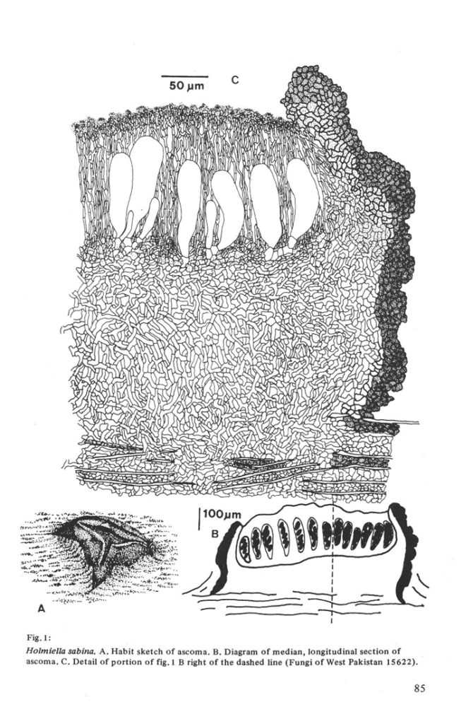
Ascomata apothecial, circular to elongate, rarely triangular, 0 5—1 mm diam., solitary to gregarious m groups of a few, at first pulvmate and covered with a continuous outer layer, outer layer splitting irregularly into 3—4 ± triangular lobes that fold back to expose a black hymemum, produced entirely withm tissue of bark, breaking through surface of bark, no stromal formation. Hymemum black when fresh and when dry. Receptacle black, smooth, slightly shining Ascomata sessile. In 3% KOH no soluble pigment or sometimes a red pigment seen.
Asci bitunicate, 4-8-spored, (85 —)105 —125(—135) x 30-45 /im, clavate to broadly clavate, base abruptly truncated, apex broad, wall thickened, with a broad
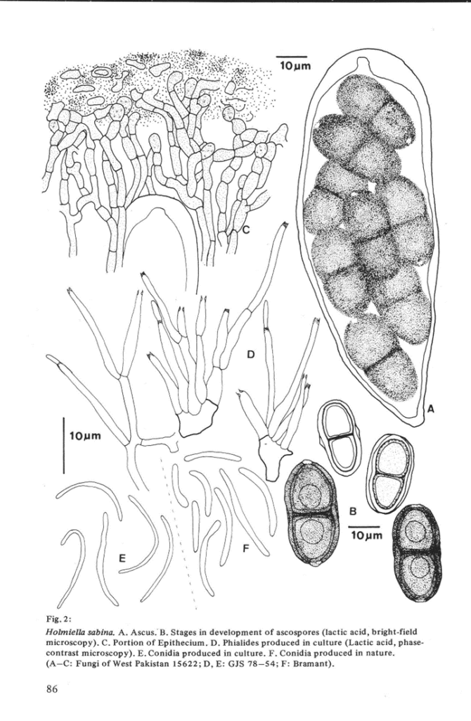
„nasse apicale”, ascal wall and apical apparatus J—. Ascospores dark brown to black, opaque, (25—)29—37(41) x (11—)13 —18(—22) jum, bicellular, with a pore m the septum, equally 2-celled or one cell slightly longer, a single drop m each cell, elliptic, not constricted at the septum, each end of each ascospore poroid (see notes below), surface of spore minutely pitted. Germinating withm 5 hrs, producing a single, unbranched, 15—25 /Um long germ tube from each end of each ascospore. Paraphyses 25—50 /im longer than asci, branching and anastomosing frequently above to form a reticulum, branching less frequently below, septate, 3—3.5 ßm wide, lightly brown pigmented, slightly spinulose above, filiform, short- celled above with tip cells subglobose, 4—5 ßm diam., becoming red m 3% KOH and pale brown m 100% lactic acid, reaction not reversible. Tips of paraphyses embedded m a continuous layer of black, anamorphous substance which turns green m 3% KOH and brown m 100% lactic acid, reaction reversible.
Subhymemum formed of bases of paraphyses and larger, pseudoparenchymatous ascogenous cells, immediately below ascal bases a thin, dense region of ± horizontally oriented, branching, hyphal cells, subhymemum lightly brown pigmented. Medullary excipulum comprised of loosly intertwined, branching, ± vertically oriented, septate, 3—4 gm wide hyphae, walls slightly pigmented, smooth, subhyahne, arising directly from the cells growing m the cortex Toward the outer edge of the ascomata cells becoming compacted, textura epidermoidea. Margin of the disc comprised of prosenchymatous, light brown cells outermost 10—15 ßm of the ascomatal wall very heavily pigmented and walls thickened.
Characteristics in culture.
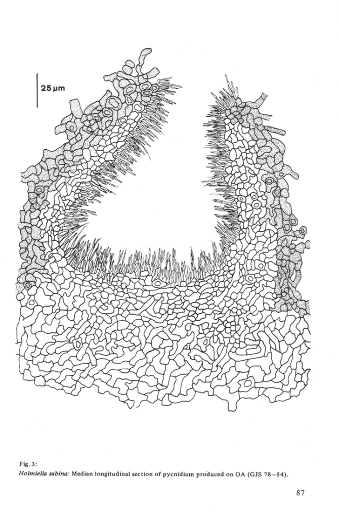
Colony characters Colonies on OA, CM, CMD, PDYE 1—2 5 (OA) cm diam, no aerial mycelium, submerged mycelium black. Comdiomata pycmdial, globose, ca 200 jum diam, black, superficial or immersed, on OA, CM and CMD forming m caespitose groups of from two to several, on PDYE the entire surface of the colony raised and forming pycmdia m a continuous crust. Pycmdia opening by splitting of the upper surface, wall ca 50 ßm wide, composed of textura epidermoidea, cells of outer ca 25 ßm heavily pigmented. Phialides cylindrical to sub-cylindrical, 10 — 17 ßm long, tapering from 1.5—2 jum wide basally to 1 — 1.5 ßm wide at the unflared opening, solitary or m pairs, arising directly from cells of the wall or terminally and laterally from short comdiophores, comdiogenous cells arising from the entire inner wall of the comdioma. Comdia stylosporous, 15 — 19 x 0.5 — 1 ßm, straight to sharply curved, unicellular, hyaline, extruded from pycmdia m hyaline slime.
Habitat.Ascomata and comdiomata found on branches of Juniperus bermudiana L., J. communis L,,J. macropoda Boiss., /. oxycedrus L., J. sabina L., /. scopulorum Sarg., J. virgimana L., and from tissues of needles and wood of J. communis. Illustrations. Müller & von Arx (1962, fig. 91 as Eutryblidiella), Holm & Holm (1977, fig. 5a, lis Eutryblidiella) Pirozynski & Reid (1966, figs. 1 — 10, as Eutryblidiella),Rehm (1896, p. 283, figs. 1—5, as Caldesia)
Specimens examined. WEST PAKISTAN: Ziarat, on branches of Juniperus sp., S. Ahmad, 26.2.1962 (Fungi of West Pakistan 15622, ZT). FRANCE: Hautes Alpes, Aguilles (Val Queyras), on Juniperus Sabina, Scheinpflug, 25.5.1958 (ZT); Hautes Alpes, Forsthaus Les Sauvas ob Montmaur, on Juniperus communis, H. Kern, 24.4.1952 (ZT); Hautes Alpes, oberes Durancetal, Argentieres, on Juniperus Sabina, E. Muller, 23.6.1958 (ZT); Savoie, Bramans, Maurienne, on Juniperus communis, E. Müller, 28.6.1966 (ZT); Savoie, oberhalb Lanslevillard, on Juniperus communis, Egger, 1.7.1966 (ZT); Var, Massif de la Ste. Baume, Hotel Miremonts, on Juniperus oxycedrus, E.Muller, 7.6.1966 (ZT); Vaucluse, Foret de St. Lambert, on Juniperus oxycedrus, E.Muller, 25.5.1962 (ZT). SWEDEN: Dolby Parish, Jerusalem, Uppland, Uppsala-Nas, on Juniperus communis, O.Petnni & K.Holm, 28.8.1978 (GJS 78—54: PDD, ZT). USA: Kansas, Rooks Co., Rockport, on Juniperus virginiana, Bartholomew 1292, 7.12.1893 (Holotype Diplodia kansensis, FH).
Notes: The ascosporal wall of H.sabina is composed of at least four layers with an epispore and endospore, each of two layers. The two layers of the epispore are usually evident at the septum. Pigmentation is first evident m the inner layers of the wall while the epispore is still hyaline. As the spore matures, the epispore also becomes brown, The ends of the spore remain hyaline and thinwalled, often the ends are slightly outwardly bulged to form a colorless cap. The outermost layer of the endospore, immediately below the lightly colored epispore, appears very thin and partially disintegrated, this development occurs as the spore matures since the wall of young spores is entire. Germ-tubes emerge only through the ends of the spore and the spore wall surrounding the emergent tube has a ragged, torn appearance suggesting that the ends of the spore are thm-walled rather than being pierced by a pore that is open to the exterior. It is often very difficult to see the poroid regions of the spores and this can be explained if these regions develop concomittantly with spore
gertmination.
Pycnidia were found on one of the specimens cited above (FRANCE: Savoie, Bramans). They appeared as small, barely erumpent, hemispherical, black spots that split irregularly to expose a disc. Pycnidia in longitudinal section are cUpulate, ca. 260 |Um wide x ca. 200 pm high. The pycnidial wall is ca. 30 pm wide, pigmented throughout and composed of cells nearly circular in outline, 7—10 pm in diameter with walls ca. 2 pm wide. Phialides were identical to these produced in culture but the conidia were shorter and broader, 11-14 x 0.5-1 pm. Anamorph:(= imperfect state) Cormculariella Karsten emend DiCosmo. Teleomorph:(= perfect state)


