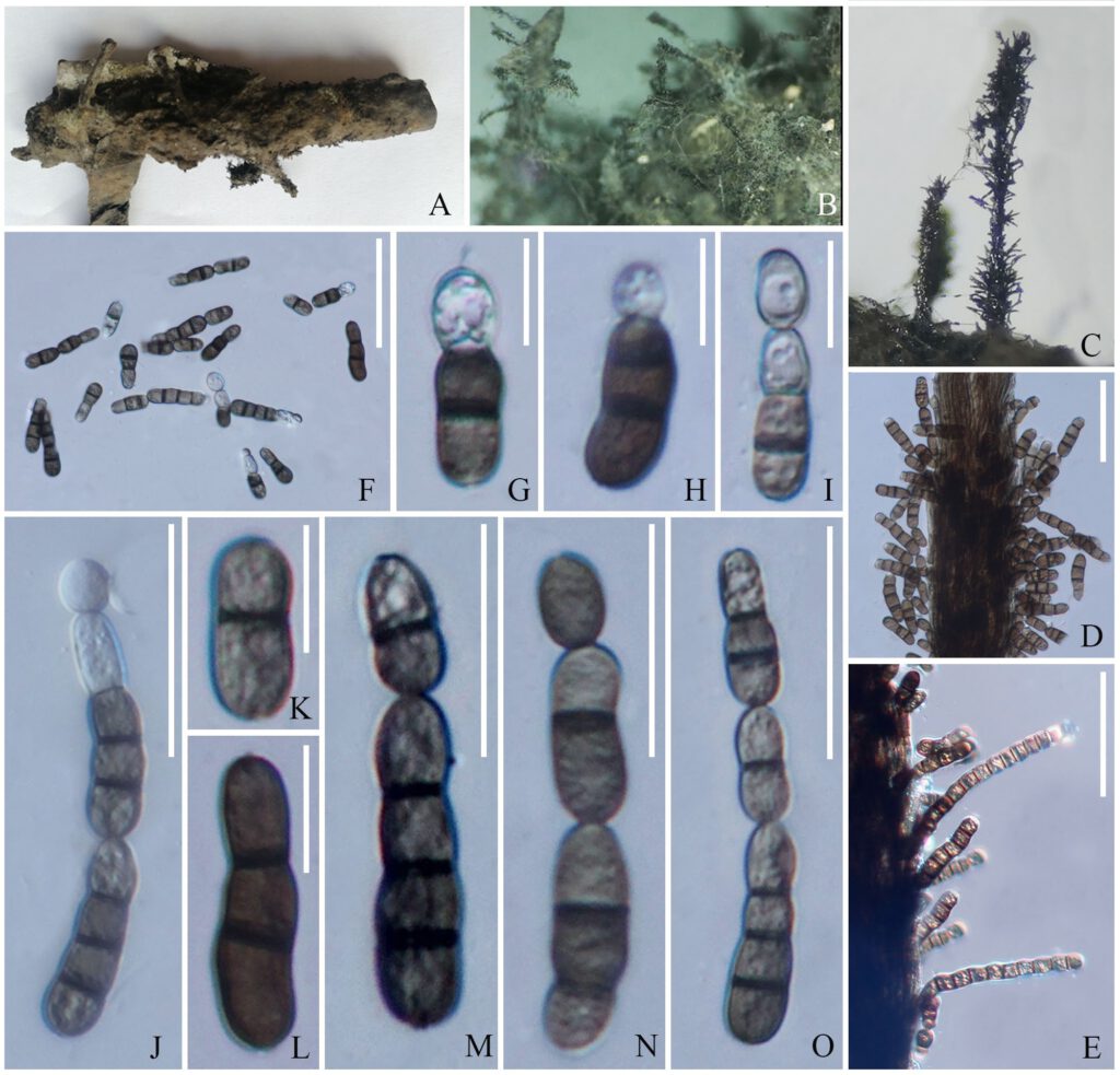
Heveicola xishuangbannaensis R.F. Xu, K.D. Hyde & Tibpromma, sp. nov. Fig 2
MycoBank number : MB 558799; Index Fungorum number: IF 558799; Facesoffungi number: FoF 10491;
Holotype: HKAS 115759
Etymology: named after the location Xishuangbanna where the fungus was first discovered.
Saprobic on rubber latex of Hevea brasiliensis. Sexual morph: Undetermined. Asexual morph: Hyphomycetous. Colonies on natural substrate consisting of long, effuse, dark, flexuous conidiophores, scattered, somewhat hairy, mass of spores all over the surface. Conidiophores 90–135 × 850–1000 μm (x̅=112.44 × 943.03 μm, n=6), aggregated synematous, macronematous, straight or slightly flexuous, pale brown, dark brown at central, septate, unbranched, smooth-walled. Conidiogenous cells reduce to conidia. Conidia 5–8 × 15–40 μm (x̅=6.53 × 26.55 μm, n=20), catenate, ellipsoidal or oblong, 0–4 to multi septate (on PDA, conidia up to 20-septate), slightly constricted at medium septum, rounded at the both end, formed in acropetal chains, initially hyaline, pale brown to brown, often with a dark brown band at the septum, branch, guttules, thick and rough-walled.
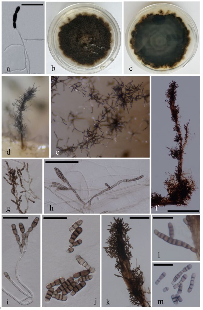
Culture characteristics: culture on PDA, colonies slow growing, umbonate, curled, smooth, edges brown, dark brown, mycelia 2.5–6 μm wide, hyaline, septate, conidia 5–12 × 10–70 μm (x̅=6.82 × 27.69 μm, n=30), oblong, up to 20-septa, pale brown to brown, thick and rough-walled (Fig. 3).
Material examined: China, Yunnan Province, Xishuangbanna Tropical Botanical Garden, on rubber latex of Hevea brasiliensis Müll.Arg., 24 November 2020, Ruifang Xu, XSBNR-07, (HKAS 115759, holotype); ex-type living cultures KUMCC 21-0086; ibid. (KUMCC 21-0086, ex-isotype).
Notes: Pin the phylogenetic analyses Heveicola xishuangbannaensis forms a monophyletic branch within Wiesneriomycetaceae with strong statistical support (Fig 1). Our new isolate is similar to Speiropsis and Phalangispora in the shape and colour of the conidia, but it can also differ in having a dark brown band at the septum and branches. While, no sexual morph has been reported so far in Wiesneriomycetaceae.

FIGURE 2. Heveicola xishuangbannaensis (HKAS 115759, holotype). A–C. Appearance of colonies on the substrate. D, E. Conidiophores with conidia. F–O. Conidia. Scale bars: D–F = 50 μm, G-I, K, L = 10 μm, J, M–O = 30 μm.

FIGURE 3. Heveicola xishuangbannaensis (KUMCC 21-0086, extype). a. Germinated conidium. b, c. Upper and lower sides of cultures on PDA. d–f, k. Sporulation on PDA medium. g–i. Conidia growing from mycelia. l Conidiophores with conidia. j, m Conidia. Scale bars: a, g = 50 μm, f = 500 μm, h = 100 μm, i = 40 μm, j, l, m = 30 μm, k = 150 μm.
